Bioenergetics: Molecular Biology, Biochemistry, and Pathology [1 ed.] 978-1-4684-5837-4, 978-1-4684-5835-0
The emergence of the Biochemical Sciences is underlined by the FAOB symposium in Seoul and highlighted by this Satellite
241 66 42MB
English Pages 484 [467] Year 1991
Front Matter....Pages i-xii
Front Matter....Pages 1-1
Introductory Remarks....Pages 3-4
Opening Lecture....Pages 5-7
Front Matter....Pages 9-9
Memories of My Flavoprotein Research....Pages 11-22
Front Matter....Pages 23-23
An Overview of Ubiquinone Proteins....Pages 25-38
Studies on the Mechanism of Action of the Mitochondrial Energy-Linked Nicotinamide Nucleotide Transhydrogenase....Pages 39-48
Structures of Beef Mitochondrial Complex I Subunits....Pages 49-55
Inhibition of the Mitochondrial Complex I Activity by Fatty Acid Derivatives of Vanillylamide....Pages 57-63
New 4-Hydroxypyridine and 4-Hydroxyquinoline Derivatives as Inhibitors of NADH-Ubiquinone Oxidoreductase in the Respiratory Chain II....Pages 65-81
Effects of Redox State for Ubiquinone-10 on the Membrane Fluidity of Cardiolipin-Containing Liposomes....Pages 83-88
Features of Assembly and Mechanism of Yeast Mitochondrial Ubiquinol:Cytochrome C Oxidoreductase....Pages 89-109
Structural Studies of Euglena Cytochrome C 1 ....Pages 111-123
How Well is Cytochrome c Engineered?....Pages 125-145
Subunit I is the Catalytic Center of P. Denitrificans Cytochrome C Oxidase....Pages 147-154
The Separation between Cytochrome A and Cytochrome A 3 in the Absolute Spectrum....Pages 155-159
Which Electron-Transferring Reactions in the Respiratory Chain Contribute to the Energy Conservation?....Pages 163-170
Cytochrome Oxidase and Peroxide Metabolism....Pages 171-180
Do Hydrogen Ions Serve as Bioenergetic Messengers in Yeast?....Pages 181-190
Molecular Organization and Regulation of the Protonmotive System of Mammalian ATP Synthase....Pages 191-204
Situation of Archaebacterial ATPase among Ion-Translocating ATPases....Pages 205-216
Structure and Chemical Modification of Pig Gastric (H + +K + )-ATPase....Pages 217-225
Front Matter....Pages 23-23
Inhibition of the Uncoupler-Induced Mitochondrial ATP-Hydrolysis by the Cooperative Work of the ATPase Inhibitor, 9K Protein and 15K Protein....Pages 227-232
Reconstitution of H + ATPase into Planar Phospholipid Bilayers and Its Kinetic Analysis....Pages 233-240
A Mitochondrial Carrier Family for Solute Transport....Pages 241-249
The Calcium Pump of the Plasma Membrane: Structure/Function Relationships....Pages 251-258
The 9-kDa Polypeptide with Iron-Sulfur Centers A/B in Spinach Photosystem I with Special Reference to Its Structure and Topographic Consideration in Thylakoid Membrane....Pages 261-270
A Photo-Signal Transducing Photoreceptor (Stentorin) in Stentor coeruleus : A Brief Review....Pages 271-274
Theory and Applications of the Kinetics of Substrate Reaction during Irreversible Modification of Enzyme Activity....Pages 275-284
High Pressure Kinetic Studies on Certain Hemoproteins....Pages 285-293
The Purification and Properties of Glycerol-3-Phosphate Dehydrogenase in the Mitochondrial Inner Membrane....Pages 295-302
Front Matter....Pages 303-303
Subunit 8 of Yeast Mitochondrial ATP Synthase: Biochemical Genetics and Membrane Assembly....Pages 305-325
Gene Structure of Human ATP Synthase Beta Subunit....Pages 327-340
Molecule and Gene of Sulfolobus acidocaldarius ATPase....Pages 341-351
Nuclear Genes Encoding Two Subunits of Human Mitochondrial Cytochrome bc 1 Complex, Cytochrome c 1 and Ubiquinone-Binding Protein: Their Structural Organization of the 5′-Flanking Regions and Chromosomal Localization....Pages 353-362
Studies on the Function of the Mitochondrial Hinge Protein: Molecular Genetic Approach Using Yeast as a Model System....Pages 363-372
Molecular evolution and biology of human mitochondrial DNA....Pages 373-387
Front Matter....Pages 389-389
Stable and Unstable Bioenergetics in Vivo ....Pages 391-398
Regulation of Activity and Tissue-Specific Expression of Cytochrome C Oxidase Genes under Normal and Pathological Conditions....Pages 399-412
Mitochondrial DNA Mutations as an Etiology of Human Degenerative Diseases....Pages 413-427
Mitochondrial Myopathies: Morphological Approach to Molecular Abnormalities....Pages 429-439
S 1 Nuclease Analysis and Direct Sequencing of Deleted Mitochondrial DNA in Myopathic Patients: Role of Directly Repeated Sequences in Deletion....Pages 441-449
Front Matter....Pages 389-389
Thirty Years of Mitochondrial Pathophysiology: From Luft’s Disease to Oxygen Toxicity....Pages 451-465
Back Matter....Pages 467-484
Recommend Papers


![Dictionary of Biochemistry and Molecular Biology [2nd ed]
0471840890, 9780471840893](https://ebin.pub/img/200x200/dictionary-of-biochemistry-and-molecular-biology-2nd-ed-0471840890-9780471840893.jpg)
![Biochemistry and Molecular Biology Compendium [1 ed.]
1420043471, 9781420043471](https://ebin.pub/img/200x200/biochemistry-and-molecular-biology-compendium-1nbsped-1420043471-9781420043471.jpg)

![Insect Molecular Biology and Biochemistry [1 ed.]
0123847478, 9780123847478](https://ebin.pub/img/200x200/insect-molecular-biology-and-biochemistry-1nbsped-0123847478-9780123847478.jpg)

![Oxford Dictionary of Biochemistry and Molecular Biology [Revised]
0198506732, 9780198506737](https://ebin.pub/img/200x200/oxford-dictionary-of-biochemistry-and-molecular-biology-revised-0198506732-9780198506737.jpg)
![Bioenergetics: Molecular Biology, Biochemistry, and Pathology [1 ed.]
978-1-4684-5837-4, 978-1-4684-5835-0](https://ebin.pub/img/200x200/bioenergetics-molecular-biology-biochemistry-and-pathology-1nbsped-978-1-4684-5837-4-978-1-4684-5835-0.jpg)
- Author / Uploaded
- Chong H. Kim (auth.)
- Chong H. Kim
- Takayuki Ozawa (eds.)
- Similar Topics
- Biology
- Biochemistry
File loading please wait...
Citation preview
BIOENERGETICS Molecular Biology, Biochemistry, and Pathology
IIOE N ERGETICS Molecular Biology, Biochemistry, and Pathology Edited by
Chong H. Kim Rensselaer Polytechnic Institute Troy, New York
and
Takayuki Ozawa University of Nagoya Nagoya, Japan
PLENUM PRESS • NEW YORK AND LONDON
Library of Congress Cataloging-in-Publication Data
Bioenergetics molecular biology, biochemistry, and pathology I edited by Chong H, Kim and Takayuki Ozawa. p. cm. Papers presented at the International Symposium on Bioenergetics, Molecular Biology, Biochemistry, and Pathology, held in Seoul, Korea, Agu. 18-21,1989, sponsored by International Union of Biochemistry and Ewha Woman's University. The symposium was held in honor of Prof. Kunio Yagi to commemorate his 70th birthday. Includes bibliographical references. Includes indexes. ISBN-Ll: 978-1-4684-5837-4
e-ISBN- 13: 978-1-4684-5835-0
001: 10.10071 973-1-4684-5835-0
1. Energy metabolism--Congresses. 2. Mitochondria--Congresses. 3. Mitochondrial pathology--Congresses. 1. Kim, Chong H., 1943II. Ozawa, Takayuki. III. Yagi, Kunio. IV. International Symposium on Bioenergetics, Molecular Biology, Biochemistry, and Patho logy (1989 Seou 1, Korea) V. Internat i Dna 1 Un i on of Biochemistry. VI. Ihwa Yo;a Taehakkyo. [DNLM, 1. Energy Metabol ism--congresses. 2. Mitochondria-metabolism--congresses. 3. Mitochondria--pathology--congresses. QH 603.M5B615 1989] QP176.B46 1990 574.19' 121--dc20 DNLM/DLC for Library of Congress 90-7851 CIP
Proceedings of an International Symposium on Bioenergetics: Molecular Biology, Biochemistry, and Pathology, held August 18-21, 1989, in Seoul, Republic of Korea
© 1990 Plenum Press, New York Softcover reprint of the hardcover 1st edition 1990
A Division of Plenum Publishing Corporation 233 Spring Street, New York, N.Y. 10013 All rights reserved No part of this book may be reproduced, stored in a retrieval system, or transmitted in any form or by any means, electronic, mechanical, photocopying, microfilming, recording, or otherwise, without written permission from the Publisher Printed in the United States of America
FOREWORD
The emergence of the Biochemical Sciences is underlined by the FAOB symposium in Seoul and highlighted by this Satellite meeting on the "New Bioenergetics." Classical mitochondrial electron transfer and energy coupling is now complemented by the emerging molecular biology of the respiratory chain which is studied hand in hand with the recognition of mitochondrial disease as a major and emerging study in the basic and clinical medical sciences. Thus, this symposium has achieved an important balance of the fundamental and applied aspects of bioenergetics in the modern setting of molecular biology and mitochondrial disease. At the same time, the symposium takes note not only of the emerging excellence of Biochemical Studies in the Orient and indeed in Korea itself, but also retrospectively enjoys the history of electron transport and energy conservation as represented by the triumvirate ofYagi, King and Slater. Many thanks are due Drs. Kim and Ozawa for their elegant organization of this meeting and its juxtaposition to the FAOB Congress. Britton Chance April 2, 1990
v
PREFACE
This book contains the contributed papers presented at the "International Symposium on Bioenergetics: Molecular Biology, Biochemistry and Pathology", held in Seoul, Korea, August 18-21, 1989, sponsored by International Union of Biochemistry (as ruB Symposium No.191) and Ewha Womans University, Seoul, Korea. The symposium was held in honor of Professor Kunio Yagi to commemorate his 70th birthday. Professor Yagi has not only made many scientific accomplishments in biochemistry, but also enormous contributions to the international biochemical communities such as International Union of Biochemistry and the Federation of Asian and Oceanian Biochemists. His unusual pleasant personality and ability to get many scientists together, have helped many biochemical societies of different countries to grow. In the last several years, advances in bioenergetics, in particular molecular genetics, have facilitated detailed study of the structure-functional mechanism of mitochondrial electron transfer and energy tranducing system, at the molecular level, and has been directed into the study of the molecular basis of mitochondrial disease, which is no longer a new field now. This is the very reason why the broad SUbtopics are chosen to be discussed in this symposium. The purpose of this symposium was to bring together the scientists from those various disciplines of bioenergetics field to discuss recent developments in their fields so that they can exchange new ideas and interact each other to promote collaborations. It was our intention to provide an opportunity to discuss a wide range of bioenergetics topics that has previously been all too rare in Asian biochemical communities. Hopefully, a meeting like this can be continued regularly, in Seoul, Korea or in Nagoya, Japan. The symposium was structured in a similar way to the previous "Symposium on Membrane Biochemistry and Bioenergetics", held in Rensselaerville, NY, August, 1986 (Kim et al., Plenum Press, 1987). Four lecture/discussion sessions and one poster/discussion session covered a variety of subjects from the molecular evolution of mitochondrial DNA to bioenergetic consequences of genetic disease. Drs. Y. Anraku, A. Azzi, M. H. Han, C. H. Kim, Q.-S. Lin, A. W. Linnane, T. Ozawa, S. Papa, P.S. Song, and C.-L. Tsou served as the session chairmen. All the participants were energetically involved in discussions of the subjects presented, giving us a lesson to plan a day more for the next meeting. All invited participants contributed a chapter to this volume except for a few, who could not make it with regret, due to various reasons. This symposium was made possible with fmancial support from sponsors. We are grateful to sponsors, the Symposium Committee of the International Union of Biochemistry, Ewha Womans University, Korea Science Foundation, MIWON Co. Ltd, Seoul, Korea and Institute vii
viii
Kim and Ozawa
of Applied Biochemistry, Gifu, Japan. We express our special thanks to Mr. Chae Bang Kim, President of MIWON Co. Ltd. for his generous support to this meeting without hesitation. We also thank to Dr. Young Bog Chae, Director of Korea Research Institute of Chemical Technology for his support. We wish to express our warm thanks to Professor Hea Chung Yun, Ewha Womans University, for her constant help throughout the organization of the symposium. We give thanks to Ms. Kum Hee Cho, Jung-Sun Kim, Hae-Jin Kim, Hye-Gyu Lee, Yong-Nam Roh, Sang-Hee Shin and Sung-Hee Shin, from Ewha Womans University for their efficient assistance during the meeting. We also thank Ms. Akiko Hashimoto in Nagoya, Ms. Donna Hilbert and Stacey Hood in RPI for their assistance throughout the organization of the symposium. Our special thanks extended to Mr. Michael Seaman for his assistance in editing and Ms. Laura Waelder for her word processing service for this volume. Finally, we are thankful for all the enthusiastic participants who made this symposium successful and hope that we can get together in the next meeting with further interesting results and ideas in the field of bioenergetics.
Chong H. Kim Takayuki Ozawa March 1990
CONTENTS
Opening Session Introductory Remarks ................................................................................................ C.H. Kim
3
Opening Lecture ........................................................................................................ E. C. Slater
5
Special Session Memories of My Flavoprotein Research ................................................................... K. Yagi
11
Part I. Biochemistry 1. Mitochondrial Electron Transfer An Overview of Ubiquinone Proteins ....................................................................... T.E.King
25
Studies on the Mechanism of Action of the Mitochondrial Energy-Linked ............. Nicotinamide Nucleotide Transhydrogenase M. Yamaguchi and Y. Hatefi
39
Structures of Beef Mitochondrial Complex I Subunits ............................................. S. Wakabayashi, R. Masui, H. Matsubara and Y. Hatefi
49
Inhibition of the Mitochondrial Complex I Activity by Fatty Acid .......................... Derivatives of Vanillylamide Y. Shimomura, T. Kawada, K. Tagami and M. Suzuki
57
New 4-Hydroxypyridine and 4-Hydroxyquinoline Derivatives as ........................... Inhibitors of NADH-Ubiquinone Oxidoreductase in the Respiratory Chain II K.H. Chung, K.Y. Cho, Y. Asami, N. Takahashi and S. Yoshida
65
Effects of Redox State for Ubiquinone-IO on the Membrane ................................... Fluidity of Cardiolipin-Containing Liposomes B.-j. Cheng, Z.-h. Yang, Y.-z. Dong, K.-r. Cheng and K.-c. Lin
83
ix
Contents
x
Features of Assembly and Mechanism of Yeast Mitochondrial Ubiquinol: .............. Cytochrome c Oxidoreductase J. A. Berden, P. J. Schoppink:, W. Hemrika and P. Nieboer
89
Structural Studies of Euglena Cytochrome c1 •••••••••••••••••••••••••••••••••••••••••••••••••••••••••• H. Matsubara, K. Mukai and S. Wakabayashi
111
How Well is Cytochrome c Engineered? .................................................................. E. Margoliash, A. Schejter, T. 1. Koshy, T. L. Luntz and E. A. E. Garber
125
Subunit I is the Catalytic Center of P. denitrificans Cytochrome c Oxidase ............ M. MUller and A. Azzi
147
The Separation between Cytochrome a and Cytochrome a 3 in the ........................... Absolute Spectrum T. Fujiwara, Y. Fukumori and T. Yamanaka
155
Part I. 2. Energy Coupling and Ion Transport
Which Electron-Transferring Reactions in the Respiratory Chain............................ Contribute to the Energy Conservation? E. C. Slater
163
Cytochrome Oxidase and Peroxide Metabolism ..... .............. .................................... Y. Orii
171
Do Hydrogen Ions Serve as Bioenergetic Messengers in Yeast? .............................. A. Kotyk
181
Molecular Organization and Regulation of the Protonmotive System of ................. Mammalian ATP Synthase S. Papa, F. Guerrieri and F. Zanotti
191
Situation of Archaebacterial ATPase among lon-Translocating ATPases ................. Y. Mukohata and K. Ihara
205
Structure and Chemical Modification of Pig Gastric (1f++K+)-ATPase .................... M. Maeda, S. Tamura and M. Futai
217
Inhibition of the Uncoup1er-Induced Mitochondrial ATP-Hydrolysis ...................... by the Cooperative Work of the ATPase Inhibitor, 9K Protein and 15K Protein T. Hashimoto, H. Mimura, Y. Yoshida, N. Ichikawa and K. Tagawa
227
Reconstitution of H+ ATPase into Planar Phospholipid Bilayers and its ................... Kinetic Analysis H. Hirata and E. Muneyuki
233
Contents
xi
A Mitochondrial Carrier Family for Solute Transport .............................................. M. Klingenberg
241
The Calcium Pump of the Plasma Membrane: StructureIFunction Relationships .... E. Carafoli, P. James, E. Zvaritch and E. E. Strehler
251
Part I. 3. Other Related Topics The 9-kDa Polypeptide with Iron-Sulfur Centers AlB in Spinach ............................ Photosystem I with Special Reference to its Structure and Topographic Consideration in Thylakoid Membrane H. Oh-oka, Y. Takahashi and H. Matsubara
261
A Photo-Signal Transducing Photoreceptor (Stentorin) in Stentor ........................... eoeruleus: A Brief Review P. S. Song
271
Theory and Applications of the Kinetics of Substrate Reaction during .................... Irreversible Modification of Enzyme Activity c.-L. Tsou
275
High Pressure Kinetic Studies on Certain Hemoproteins ......................................... C. Balny
285
The Purification and Properties of Glycerol-3-Phosphate Dehydrogenase .............. in the Mitochondrial Inner Membrane Q.-s. Lin, S.-p. Shi and J.-h. Liu
295
Part II. Molecular Biology Subunit 8 of Yeast Mitochondrial ATP Synthase: Biochemical Genetics ................. 305 and Membrane Assembly P. Nagley, R. J. Devenish, R. H. P. Law, R. J. Maxwell, D. Nero and A. W. Linnane Gene Structure of Human ATP Synthase Beta Subunit............................................. Y. Kagawa and S. Ohta
327
Molecule and Gene of Sulfolobus aeidoealdarius ATPase ....................................... K. Denda, J. Konishi, K. Hajiro, T. Oshima, T. Date and M. Yoshida
341
Nuclear Genes Encoding Two Subunits of Human Mitochondrial ........................... Cytochrome be) Complex, Cytochrome e) and UbiquinoneBinding Protein: Their Structural Organization of the 5 -Flanking Regions and Chromosomal Localization H. Suzuki, Y. Hosokawa, H. Toda, M. Nishikimi, A. Matsukage, M. C. Yoshida and T. Ozawa
353
1
Contents
xii
Studies on the Function of the Mitochondrial Hinge Protein: Molecular ................. Genetic Approach Using Yeast as a Model System C. H. Kim and R. S. Zitomer
363
Molecular Evolution and Biology of Human Mitochondrial DNA ..... ........ .......... ... S. Horai and K. Hayasaka
373
Part III. Mitochondrial Pathology Stable and Unstable Bioenergetics in Vivo ................................................................ B. Chance
391
.Regulation of Activity and Tissue-Specific Expression of Cytochrome c........... ..... Oxidase Genes under Normal and Pathological Conditions B. Kadenbach, U. Bilge, F.-J. Hilther, C. Thiel, M. Droste,A. Schlerf, B. Schneyder, L. Kuhn-Nentwig and J. Milller-HOcker
399
Mitochondrial DNA Mutations as an Etiology of Human ........................................ Degenerative Diseases T. Ozawa, M. Tanaka, W. Sato, K. Ohno, S. Sugiyama, M. Yoneda, T. Yamamoto, K. Hattori, S.-i. Ikebe, M. Tashiro and K. Sahashi
413
Mitochondrial Myopathies: Morphological Approach to.......................................... Molecular Abnormalities T. Sato, S. Nakamura, H. Hirawake, E. Uchida, Y. Ishigaki, K. Seki, R. Kobayashi, S. Horai, and T. Ozawa
429
Sl Nuclease Analysis and Direct Sequencing of Deleted Mitochondrial.................. DNA in Myopathic Patients: Role of Direcdy Repeated Sequences in Deletion M. Tanaka, W. Sato, K. Ohno, T. Yamamoto, and T. Ozawa
441
Thirty Years of Mitochondrial Pathophysiology: From Luft's Disease .................... to Oxygen Toxicity L. Ernster and C. -po Lee
451
Appendix Participants
469
Author Index .............................................................................................................
475
Subject Index .............................................................................................................
477
Opening Session
INTRODUCTORY REMARKS
Chong H. Kim
It is my pleasure to open the "International Symposium on Bioenergetics: Molecular Biology, Biochemistry and Pathology" in honor of Professor Kunio Yagi. I welcome all of you to Seoul, Korea! I think that most of you - probably all of you who is not resident here - come to Seoul for the first time. Before I introduce Professor E.C. Slater, President of International Union of Biochemistry, for opening lecture, I would like to say a few words for having this symposium in Seoul. It is a very exciting moment for me to have all of you here at the Symposium on Bioenergetics. Because there is a reason. It has been fifteen years since I left my homeland, Seoul, Korea, to pursue advanced studies in Biochemistry in the United States. At that time, it was very difficult to do any advanced biochemical experiments in Korea. I had already done a few rather simple biochemical experiments by that time. However, I had to run from one institute to another in order to accomplish even those simple experiments. Unfortunately, I could not find a good teacher to lead my enthusiasm towards biochemical research. Therefore, I decided to study biochemistry in the States, where I had already visited for a month in 1973, on the way of my return from Copenhagen, Denmark, where I spent a few months for the advanced courses in Clinical biochemistry. Fifteen years later now, Korean Biochemical Society has impressively grown to the level, at which they can freely exchange and discuss their scientific progress with leading scientists from all over the world. Some of you must have seen such an example at the 5th FAOB (Federation of Asian and Oceanian Biochemists) Congress, which was held last week just before this symposium. I am very proud to see that such an excellent international scientific gathering has been held in Seoul. Our decision to hold a Symposium on Bioenergetics in Seoul, Korea, came about a year ago in Prague, at the 14th International Congress of Biochemistry. It is my honor to have a "Symposium on Bioenergetics" participated by all you, leading scientists from 13 different countries, in the country I was born and educated, with the support of the institution at which I was educated. I am grateful to those of you, my teachers, friends and colleagues, who have nourished my knowledge in bioenergetics, and helped me to open this successful, I believe, symposium in Seoul. Above all, I thank my co-organizer, Professor Ozawa, who is an excellent partner. I must also mention about the person behind me to make the smooth running of this symposium, Professor Hae-Jung Yun, Dean of College of Pharmacy, Ewha Womans University. Her unaccountable help made my task for this meeting easier.
3
4
Kim
I hope all of you can have a joyful time during this meeting even though our schedule is too tight and this can be a momentum to bring another Bioenergetics meeting in Seoul near future. I especially invite you to experience the beauty of the city of Seoul at the end of this meeting, and now I would like to introduce Professor Slater for the opening lecture.
OPENING LECTURE
E. C. Slater
Thank you very much, Dr. Kim and Professor Yagi and Dr. Ozawa and to other colleagues. There are, of course, many reasons why I am happy to say a few words at the opening of this symposium. First, and foremost, it is my pleasure to be able to attend this meeting to honor Professor Yagi on the occasion of his 70th birthday. I shall have more to say about you and to you, Kunio, tomorrow evening, and it suffices now to congratulate you and welcome you to the select band of septuagenarians present at this meeting. Now another reason why it is a great pleasure for me to say a few words here is that this is a ruB symposium, it is #192 as a matter of fact and I have attended all of the IUB congresses since the fIrst one in 1949. I, of course, cannot say the same thing about IUB symposia, even during the period in which I am president of ruB. This year alone IUB is sponsoring no less than 28 symposia through its two committees-Symposium Committee which is chaired by Arnost Kotyk who is here at this meeting and our so-called Interest Groups Committee. But, although we have allocated more than one-third of our total budget - our total budget is only about $300,000 to support our symposia, a quick calculation will show you that our support is spread out pretty thinly. Actually, Dr. Kim has been most skillful in utilizing our resources effIciently. Since quite apart from direct subvention from our Symposium committee - three of the speakers are members of the executive committee of ruB who are meeting here in Seoul, earlier in the week, and have had their travel expenses paid by IUB under a different budget heading, namely of the executive committee. Moreover, others have attended the FAOB Congress which ended yesterday, which also received some support from IUB under yet another budget heading, and I am sure that our Treasurer, who is Tony Linnane, will be very pleased at your efficient use whereby money is spent twice. This is actually the second IUB sponsored symposium on bioenergetics held in the FAOB area. I suppose those who were not at this very wonderful FAOB meeting earlier this week - really extremely well organized meeting with a good program indeed, and some of you who have just flown in may not know what FAOB is - well let me tell you it is the Federation of Asian and Oceanian Biochemists. Now the fIrst ruB symposium in Asia was held in Beijing, China in 1984, in Bioenergetics. I just want to sketch because Dr. Kim asked me to fill up the program up to 9:00 o'clock. The Progress that I see or the sort of changes that I see have come into the field in the last five years - during much of the time which I have no longer been active in bioenergetics research. I think, of course, a lot of progress was made particularly by the application of techniques introduced from molecular genetics. Of course, cloning and DNA sequencing
5
6
Slater
had already, five years ago, given us the amino acid sequences of many subunits of the electron transferring and ATP-synthesizing proteins. This has been largely completed now in the intervening period. And the powerful tool of site-directed mutagenesis has been directed to the study of the function, mechanism of action and particularly in the biosynthesis also of the subunits. I think: that we can say that the mechanism of electron transfer or the pathway, in any case, of electron transfer within the large electron transferring proteins of the mitochrondiron has been largely worked out. Also, we now know rather more than we did five years ago about which electron transferring reactions are energy conserving, that is, they contribute to the charge separation across the membrane. Although I am not sure this will be discussed in this symposium - I hope it will be -. We now have a deeper insight into the forces which drive electron transfer between different well separated centers in the protein. I do not think we have progressed very far in our understanding of how an electric field across a membrane or protonmotive force, if you will, catalyses the synthesis of ATP or the dissociation of already formed ATP but I may be wrong - maybe you will prove me wrong at this meeting. Perhaps the most striking and unexpected development - unexpected for me in any case - in recent years has been in the molecular basis of mitochondrial pathology, which is the last topic that will be dealt with in this meeting. For me, my first surprise came from Dr. Kadenbach's demonstration of the tissue-specific expression of cytochrome oxidase genes. In any case, this made it more understandable why certain inborn errors may lead to lesions in specific tissues. I have also been surprised by the change of attitudes to mitochondrial disease, since I left the laboratory. Where it was once thought to be very rare, it now appears that the cases examined are the tip of an iceberg or perhaps we should say for the investigators, the exposed reef of a gold mine. It is not only the genetically inherited disorders of the respiratory chain that are attracting attention, but also those that arise owing to mutations of the mitochondrial DNA after birth. Indeed, Tony Linnane and Takayuki Ozawa have suggested that the failures in function associated with aging - which is something, that is of interest to you and I, Kunio - are due to a progressive loss of respiratory capacity of tissues as a result of random mutation of the mitochondrial DNA. I must say that it is of some comfort to know that the field, in which I have worked during most of my scientific life, might now be providing me with an explanation of why I find it more difficult to follow the work of my younger colleagues nowadays - who, of course, still have beautifully operating mitochondria producing kilograms of ATP per hour, and even the increasing number of mistakes I make in proofreading papers and letters. In the late 1940s and in the 1950s, bioenergetics belonged to the cutting edge of biochemistry. if you can excuse the jargon. New fields opened up by the discovery of the structme of nucleic acid superseded bioenergetics in the center of interest. In the middle of the 196Os, a new era opened up with the discovery that mitochondria have their own genetic apparatus distinct from that present in the nucleus. However, this did not have much itnpact outside of mitochondria - others considered it of secondary importance - a sort of evolutionary fossil, retained after bacteria established themselves as symbionts in the animal cells and evolved into mitochondria. For a short period in the 1970s bioenergetics became popular again with the granting agencies when it was suggested that the combination of photochemical reaction centers and hydogenase could be utilized on a commercial scale to provide hydrogen gas to provide fossil fuel. This is not yet possible, but the relative priority given to this study, did I believe, lead to the discovery of the role of nickel in hydrogenase and related enzymes, and that of manganese in the oxygen evolving reaction
Opening Lecture
7
of photosynthesis a little more rapidly than otherwise might have been the case, because up till then support in the very rapidly productive field of photosynthesis was declining. The concept that mitochondrial diseases are not only more important than we thought, but perhaps universal since all of us age - even you younger bioenergeticists age -, opens new possibilities and perhaps it will become a major field of medicine. To those in the field - even if not always to those outside the field-bioenergetics, has always been exciting. To paraphrase Arthur Kornberg in another context, "I have never met a dull electron transferring protein." The fundamental importance of the electron transport chain and oxidative phosphorylation is, of course, obvious to everyone. But some might have felt that the main features have long been established and have been incorporated into textbooks - just like the Krebs cycle has - and it only remains to dot the "i"s and cross the "t"s. However, the recent developments in the importance of mitochondrial disease, for example, would never have taken place if the filling in of details - the dotting the "i"s and the crossing the "t"s, particularly in the connection of the mechanisms of biogenesis of mitochondria had not been pursued. I would like to conclude this opening remark by thanking Dr. Kim and Dr. Ozawa for putting together this interesting symposium and inviting many of us. Practically everybody here visits Seoul for the frrst time. I can assure you that we are extremely impressed by what we have seen here in Seoul and how you organized the extremely successful meeting. I also would like to thank Ewha Womans University for sponsoring this meeting together with ruB and also those co-sponsors. Thank you!
Special Session
MEMORIES OF MY FLAVOPROTEIN RESEARCH
Kunio Yagi Institute of Applied Biochemistry, Yagi Memorial Park Mitake, Gifu 505-01, Japan
Friends! Ladies and Gentlemen! It was, and still is, a great pleasure and honor to me when I learned that the present symposium was to be held in honor of myself. Before continuing further, I want to express my heartfelt thanks to the members of the organizing committee and to all participants in this symposium. Generally speaking, I am a very future-oriented person and do not often focus on past events. However, on very rare occasions, such as the present one, I am prompted to do precisely that. So, during the next few minutes I want to share with you some of the historical background of my past research, especially that on flavoproteins, not failing of course to also bring in the element of human relationships. When I was a middle-school student in Tokyo, sometime around 1932 to 1936, I had aspirations of becoming a novelist or poet, and so I entered the literary program at Tokyo Metropolitan High School. During the three-year term of high school, however, I began to have second thoughts about my choice of future career. At some point I changed my mind and decided that I would become a medical researcher instead, since medicine seemed to me a more exciting endeavor than pen and ink. So, in 1936, I entered medical school. Since Tokyo University as well as Kyoto University did not allow literary students to apply, I decided to apply to the medical school at Nagoya University. Shortly after entrance there, my excitement waned considerably, especially when I attended the lectures on anatomy, a topic that seemed to me very similar to geography. In addition, I realized that I would not be able to pass the biochemistry examination, for my knowledge of chemistry was so poor. Thus, I decided to spend most of my time during the first year of medical school studying chemistry. Fortunately, the professor of biochemistry was very considerate of me and allowed me the use of a bench in his laboratory. Thereafter I conducted primitive experiments in chemistry on a daily basis until one eventful day. On that day the dean of the medical school happened to visit the laboratory and asked me, "What are you doing?" "Chemical experiments," I answered very politely. Then he asked me, "Are you a student?"; and I replied, "Yes." He then continued, "In this medical school, students must attend the lectures. What lecture is being given now?" He next inquired, Promptly I answered, "Hygiene."
Bioenergetics, Edited by C. H. Kim and T. Ozawa, Plenum Press, New York, 1990
11
Yagi
12
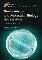
Fig.
1.
A
portrait
of
Prof .
Leonor
Michaelis.
"Why aren't you attending this lecture?" I answered with all honesty, "A lecture on hygiene does not interest me." In response he di s played a most unpleasant face and then asked me to corne to his office later. When I entered his office at the appointed time, he persuaded me to attend all of the lectures, but, to my relief and great joy, he also informed me that he was giving me official permissi on to conduct e x p eriments in the biochemical laboratory at any time, as long as it was between evening and morning! And then he added, "Night is not only for sleeping!" Thus, having been granted a position of sorts in the biochemical laboratory, I carried out experiments every night. When I was tired, I often slept in the lab. Fortunately for me and to my surprise, there in the laboratory was of all things a very beautiful, large, black-painted bed decorated with golden designs! So I gratefully put it to good use, but thought it odd that such a disharmonious thing should be present in a laboratory! One day I asked a senior staff membe r of the labo ratory about this most interesting bed. He enlightened me, saying "This was the bed of Professor Leonor Michaelis. He is the founder and was the first professor of this laboratory. As you will learn in the biochemistry course, he is the very person who proposed the MichaelisMenten theory (1). Also, you must read this book." Then he showed me a book written by Prof. Michaelis entitled "Die Wasserstoffionen-Konzentration" (2). Thus, this was my first contact with Prof. Michaeli s . Figure 1 shows a portrait of this very distinguished gentleman (3). Prof. Michaelis is, of course, famous for the theory that bears his name along with that of his colleague, in · addition to his many other pioneering works, but few people know about his c ontribution t o t he Japanese biochemical community in its early days . In 1922, Prof. Michaelis came to Japan from Germany at the invitation of Aichi Medical College, now the University of Nagoya, and was asked to found a department of medical chemistry, which is now known as the Institute of Biochemistry in the Faculty of Medicine of the University of Nagoya. Figure 2 shows a photograph of Prof. Michaelis with his family and colleagues in front of the college (4).
13
Memories of Flavoprotein Research
Fig. 2. Prof. his family and
Leonor Michaelis (fourth from the left) colleagues in front of the college.
with
In 1923, one year after his arrival, he began lectures for medical students; and, in addition, he began conducting some highly inspiring and stimulating special courses for young Japanese biochemists in this new department. Figure 3 is a photograph depicting the Professor in the process of demonstrating an experiment to his eager class (5).
(fourth Fig. 3. Prof. Leonor Michaelis conducting a physico-chemical experiment.
from
the
left),
Yagi
14
Fig. acid
4.
Crystals of the From ref. 9,
oxidase.
enzyme-substrate complex of D-amino of Elsevier Publishing Company.
courtesy
When I was studying as a medical student in the Department of Biochemistry, School of Medicine, Nagoya University, during the period 1939-42, some years had passed since Prof. Michaelis had left Nagoya, but many things he had brought from Germany still remained and somehow strongly influenced me. In addition, when I started my research work just after the Second World War, the mechanism of enzyme action was one of the most challenging targets for young biochemists. Feeling somewhat close to Prof. Michaelis and excited by his now famous theory, I decided to study the enzyme-substrate complexes (6). Prof. Michaelis had predicted a single enzyme-substrate complex, that is, the Michaelis complex, to exist during an enzymatic reaction: E
+
s
E
s
E
+
P
However, many people thought later that a series of intermediate complexes could exist in the reaction sequence: E
+
s セ@
E
s
E
p セ@
E
+
p
While contemplating just how to demonstrate such intermediate or intermediates, I thought that an enzyme having characteristic properties such as a color, as is the case for flavin enzymes, would be advantageous. I finally selected D-amino acid oxidase, a dissociable flavin enzyme, as a model enzyme, since I presumed that the flavin chromophore would serve as an indicator for the oxidation/reduction state, as well as for the formation of any complex. As a working hypothesis, I reasoned that if an enzyme were to form an enzyme-substrate complex, which would then result in the enzyme and product, and if the enzymatic reaction were reversible, then a certain amount of the complex should become detectable in the presence of adequate amounts of the substrate and product. Furthermore, I assumed that the complex might become stabilized under reaction conditions other than those giving the maximal reaction and that the complex might thus be crystallizable under some appropriate conditions (7). With these thoughts in mind, I carried out extensive research, and after much hard work of long duration, I succeeded, in collaboration with Dr. Takayuki Ozawa, in the crystallization of a beautiful purple-colored enzyme-substrate complex, which
Memories 0/ Flavoprotein Research
15
1.2
Q)
u s::
'"o 0.6 1: Vl
.0
cI:
Wavelength (nm) Fig. 5. oxidase.
Identity Changes
of in
the
the
purp1e
absorption
intermediate spectrum of
of
D-amino
acid
D-amino acid oxidase
after the addition of D-alanine under the anaerobic condition. a, Oxidized enzyme; b, 300 msec after mixing; c, 2 sec after mixing. From ref.
17,
courtesy of
the
Japanese Biochemical
Society.
was composed of equimolar amounts of D-amino acid oxidase and the substrate D-alanine. Figure 4 shows the crystals of this complex, which, incidentally, was the very first enzyme-substrate complex to be crystallized. The year was 1962. We submitted a short paper describing this exciting finding to Biochimica et Biophysica Acta (8). Professor E. C. Slater, who is among us tonight, accepted our paper for publication in this journal after he had kindly edited the manuscript in his own handwriting. After the pUblication of this paper, which was followed by a series of related communications (9-12), I rapidly became recognized by many leading biochemists and was given many opportunities to present our work at various symposia around the world. At one such symposium, "Mechanism of Enzyme Action," at the 5th Congress of the International Union of Biochemistry, held in New York in 1964, my presentation was reported by Chemical and Engineering News (13). Their report resulted in even wider recognition of our findings. However, opposition soon arose. When I spoke at the First Symposium on Flavins and Flavoproteins, organized by Prof. Slater and held in Amsterdam in 1965 (14), a serious question about our results was raised by Dr. Vincent Massey (15), who, incidentally, is also present here tonight. By the stopped-flow technique, he and his colleagues (16) had measured the absorption spectrum of the purple-colored intermediate that appeared during the reaction of D-amino acid oxidase with D-alanine under the anaerobic condition. By their procedure they discovered that the spectrum measured at 150 ms after the anaerobic mixing of the enzyme and substrate differed from the one measured at 1.8 s after the mixing. As the spectrum of our crystals was different from theirs measured at 150 ms, but identical to theirs measured at 1.8 s, they therefore argued that our crystallized species was meaningless in the catalytic sense because a 1.8-s species could not explain the high molecular activity of this enzyme.
Yagi
16
e
c 1.5
1.0
0.5
ᄚPセMTUV@ Wavel ength (nm) Fig. 6. Changes in the absorption intermediate complex during its aging Purple after
intermediate 10
days;
Publishing
e,
complex;
after
1
b,
month.
after From
3
spectrum of in the dark days;
ref.
10,
c,
after
courtesy
the purple at 5°. a, 5 of
days;
d,
Elsevier
Company.
When we looked carefully at their results, we found them to be correct and quite reproducible. Figure 5 shows the data we obtained on this point by rapid-scanning spectroscopy (17). These new findings naturally had a strong impact on me, and so we set out to examine this problem extensively. When we obtained the absorption spectrum of the "rapidly appearing intermediate" and that of the "slowly appearing one" and compared the two, we found that the difference spectrum between them agreed with that between the oxidized enzyme and the "slowly appearing intermediate". We viewed this phenomenon as indicating that the "rapidly appearing intermediate" was not homogeneous but rather was consisted mostly of the purple intermediate and, to a minor extent, of the oxidized enzyme. This indicates that most of the enzyme changes rapidly to the purple entity, whereas a lesser portion does so only slowly. As we were already aware of the monomer/dimer equilibrium of Damino acid oxidase (18,19), we interpreted these data to indicate that these two forms of the enzyme were responsible for these two entities; that is, the dimeric form reacts more rapidly with the substrate to form the purple intermediate than does the monomeric form of the enzyme. The quantitative analysis supported this assignment (17). Therefore, we concluded that the crystalline purple intermediate is, in fact, the intermediate appearing during the enzyme catalysis. This different catalytic activity between monomer and dimer of Damino acid oxidase generated additional interest in the monomer/dimer equilibrium of enzymes in general. Furthermore, this finding provides a warning that the adjustment of parameter values obtained with a low enzyme concentration by those obtained with a high concentration of
Memories of Flavoprotein Research
Fig. 7. oxidase.
Crystals
of
From
11,
ref.
the
17
semiquinoid
c ourtesy
of
El se vier
form
of
D-amino
Publi shi ng
acid
Company.
enzyme is crucial, because a low concentration of enzyme contains a larger amount of monomer than does a high concentration of enzyme, as evidenced by D-amino acid oxidase (20,21). Another question that arose from the crystallization of this enzyme-substrate complex was the nature of the complex itself. Judging from the purple color of the crystals, I thought initially th a t the coenzyme portion of the entity would be in the semiquinoid form, especially since it was already established that the oxidized form of the enzyme has a yellow color whereas the fully reduced form is colorless. To e xamine this possibility, we conducted an elec t ron-spin res o nance study on the crys tal. Although we did detect a signal (22), quantitative analysis revealed that only a minute portion of the coenzyme, flavin adenine dinucleotide (FAD), was a radical species. Additionally, the amount of radical contained in the crystals varied with time. When we looked into the matter more closely, we found that the crystalline complex initially h a d no signal but that the signal gradually appeared and increased, with th e spectrum of the complex gradually changing from that of a purple to that of a red species (10). Figure 6 depicts th e spectral changes. These findings indicate the initial complex to be a diamagnetic species and the gradually transformed one to be a paramagnetic one. By exhaustive examination of the phenomenon we finally concluded that the complex is a strong charge-transfer complex that dissociates gradually into radical cation and radical anion (10,23). The substrate radicals disproportionate to yield non-radical species, whereas the flavin radicals remain unchanged owing to their stabilization by the apoprotein. By starting with the purple complex, we were able to crystallize the semiquinoid enzyme of D-amino acid oxidase (11). Figure 7 shows such crystals, the first ever prepared of a semiquinoid flavin enzyme. Since the purple intermediate complex was found to be the very one that reacts with oxygen (24), we assumed that the slow dissoci a tion of the complex, that is, the long life time of this strong charge-t ra nsfer complex, is responsible for gi v ing the complex sufficient time t o react with the oxygen. The reason for this complex being somewhat stabili zed
Yagi
18
QJ
U
co
QJ
U
VI QJ
S-
o
::J
;:;::
0
450
550
500 Wavelength (nm)
of the spectrum fJ.uorescence Fig. S. Changes in the apoenzyme-MBAS compJ.ex upon the addition of FAD. a, Apoenzyme; b, MBAS (92 J.LM) ; c, a + b; d, c + FAD (21.8 J.LM) ; e, c + FAD (43 J.LM); f, nm.
c
+ FAD
(108
From ref.
J.LM);
26,
g,
reconstructed
courtesy of the
holoenzyme.
Excitation
at
362
Japanese Biochemical Society.
became the next problem for us to tackle. We predicted that the microenvironment surrounding the charge-transfer interaction might be hydrophobic in nature. To test our prediction, we synthesized several hydrophobic probes that fluoresce when they combine with hydrophobic regions of proteins (25). Among these probes, we found the novel compound monobenzoylated 4,4'-diaminostilbene 2,2'-disulfonate (MBAS) to be suitable, for this dye can substitute for the coenzyme FAD and combine with the apoenzyme of D-amino acid oxidase (26). As shown in Figure 8, when the dye combines with the apoenzyme, the former fluoresces; and this fluorescence diminishes upon the addition of FAD. The fluorescence spectrum finally becomes identical to that of the D-amino acid oxidase holoenzyme, indicating that the coenzyme resides in a hydrophobic pocket of the apoprotein (26). Additionally, we found that another hydrophobic probe, 1-anilinonaphthalene-8-sulfonate, can combine with the apoenzyme in competition with the substrate D-alanine (26) . Therefore, we concluded the microenvironment surrounding the charge-transfer interaction to be hydrophobic. I presented these results just mentioned at the Second International Symposium on Oxidases and Related Redox Systems, which was held in Memphis in 1971 (27). I will never forget the witty comment made by Dr. W. E. Blumberg (28) after my presentation. He said, "In case anybody missed the point of Dr. Yagi's talk in relevance to the discussion earlier today, let me reiterate that one could paraphrase and say that anybody who is talking about flavin water chemistry in this enzyme is all wet!" From another point of view, such a hydrophobic environment for the coenzyme was also found to be important for the catalytic activity of the enzyme, for our molecular orbital calculations demonstrated the
19
Memories of Flnvoprotein Research
/
O-H
H
Fig. I,
9.
II,
Geometry
III,
courtesy
of
of
IV,
and
the
Japanese
oxidized
V indicate
lumiflavin-water
hydrogen-bonding
sites.
complexes. From
ref.
31,
Biochemica.l Society.
importance of hydrogen bonding at particular heteroatoms of the isoalloxazine nucleus of flavin for its reactivity (29-31). Figure 9 shows the geometry of various oxidized flavin-water complexes. I-V indicate sites of hydrogen bonding. In the oxidized flavin, the N(5) atom most probably accepts electrons (32). The atomic orbital coefficients in the lowest unoccupied molecular orbital at N(5) of oxidized lumiflavinwater complexes indicated that hydrogen bonding at all heteroatoms
H
H
-::"·1 o
H
H, H"".,.
H 1111". C
H'"
Fig. I,
I I,
10. III,
courtesy
of
セ@
H
H
Geometry IV,
of
and V
Elsevier
reduced
indicate
Publishing
lumiflavin-water
hydrogen-bonding Company.
sites.
complexes. From ref.
30,
20
Yagi
except N(3) increases the electron acceptability of N(5). Figure 10 shows the geometry of reduced flavin-water complexes. In the reduced flavin, C (4a) is the atom that donates electrons to a neutral electrophile such as oxygen (33). The atomic orbital coefficients in the highest occupied molecular orbital at C(4a) of reduced lumiflavin-water complexes indicated that hydrogen bonding at N(l) or N(3) increases the electron-donating ability of C (4a) . If the microenvironment of the coenzyme were aqueous, all heteroatoms would be expected to form hydrogen bonds with water molecules, and such hydrogen bonding at particular heteroatoms would be impossible. However, hydrogen bonding between selected heteroatoms of flavin and amino acid residues in a hydrophobic region of the apoenzyme would explain the increased reactivity of the coenzyme noted when flavin combines with the apoenzyme. Such hydrogen bonding in a hydrophobic environment answers, at least in part, the question raised by Professor Hugo Theorell, who, incidentally, invited me to collaborate with him in his laboratory back in 1957. In his Nobel Lecture (34) in 1955 he said, "We still do not quite understand how through its linkage to the coenzyme the enzyme-protein 'activates' the latter to a rapid absorption and giving off of hydrogen." Before concluding my lecture, I would like to mention some of the human aspects related to the work I have just presented to you. Professor Slater, who accepted our paper in his capacity as an editor of Biochimica et Biophysica Acta, came to Nagoya in 1973 at my invitation, and attended the symposium that we dedicated to the late Prof. Michaelis on the occasion of the 50th anniversary of the commencement of his lectures in Nagoya. In that meeting, Prof. Slater gave a warm address (35) that afforded considerable pleasure to those colleagues of Prof. Michaelis who were present at that time, though they have all passed away by now. Professor Massey, who had argued initially about the purple crystals, became a very good friend of mine thereafter; in fact, in 1985 he even collaborated with me at my institute in Japan. Doctor Ozawa was a post-graduate student at the time of the crystallization of the enzyme-substrate complex. Today he is a Full Professor of Biochemistry at the University of Nagoya, and is one of the organizers of this symposium. I view science as a human enterprise that has no end, whereas the scientist himself or herself, during an all too brief period of time, adds his or her contribution and then is no more. During the plying of our trade, we make personal contact with a multitude of like persons: we compete, we argue, we cooperate, and we even become intimate friends. Truly we are a lucky or fortunate segment of society. This present symposium is yet another example of such interactions among us scientists! Ladies and Gentlemen! Thank you once again for your kind attendance, and I wish each of you much future success and good luck!
ACKNOWLEDGEMENT The major portion of this paper was reproduced, in paraphrased form, from the following article by the same author that appeared in Biochim. Biophys. Acta 1000 (1989) 203-206: Commentary by Kunio Yagi on 'Crystallization of Michaelis complex of D-amino acid oxidase' by K. Yagi and T. Ozawa, Biochim. Biophys. Acta 60 (1962) 200-201. The author thanks Elsevier Publishing Company for its permission to reproduce this information.
Memories of Flavoprotein Research
21
REFERENCES
1. Michaelis, L. and Menten, M. L. (1913) Biochem. Z. 49, 333-369 2. Michaelis, L. (1922) Die Wasserstoffionen-Konzentration, SpringerVerlag, Berlin 3. This portrait of the late Prof. Michaelis was kindly furnished by Prof. Michaelis's daughters, Mrs. llse Michaelis Wollman and Mrs. Eva Michaelis Jacoby, upon their coming to Japan in 1973 on the occasion of the symposium dedicated to the late Prof. Michaelis in commemoration of the 50th anniversary of the opening of his lecture in Aichi Medical College 4. This picture was kindly furnished by the late Prof. I. Ogawa of the University of Nagoya, who was a staff member of Prof. Michaelis 5. This picture was also donated by the late Prof. I. Ogawa 6. This story was described in Reactivity of Flavins (The proceedings of the symposium dedicated to the late Professor Leonor Michaelis under the auspices of the Japanese Biochemical Society) (Yagi, K. ed.), Univ. Tokyo Press, Tokyo, 1975 7. Yagi, K. (1965) in: Advances in Enzymology (Nord, F. F. ed.), Vol. 27, pp 1-36, lnterscince, New York 8. Yagi, K. and Ozawa, T. (1962) Biochim. Biophys. Acta 60, 200-201 9. Yagi, K. and Ozawa, T. (1964) Biochim. Biophys. Acta 81, 29-38 10. Yagi, K., Okamura, K., Naoi, M., Sugiura, N. and Kotaki, A. (1967) Biochim. Biophys. Acta 146, 77-90 II. Yagi, K. , Sugiura, N. , Okamura, K. and Kotaki, A. (1968 ) Biochim. Biophys. Acta 151, 343-352 12. Yagi, K. , Okamura, K. , Sugiura, N. and Kotaki, A. (1968) Biochim. Biophys. Acta 159, 1-8 13. Chemical and Engineering News 10, 34, 1964 14. Yagi, K. (1966) in: Flavins and Flavoproteins (Slater, E. C. ed.), pp 210-222, Elsevier, Amsterdam 15. Massey, V. (1966) in: Flavins and Flavoprotein (Slater, E. C. ed.), p 222, Elsevier, Amsterdam 16. Massey, V., Palmer, G., Williams, C. H. Jr., Swoboda, B. E. P. and Sands, R. H. (1966) in: Flavins and Flavoproteins (Slater, E. C. ed.), pp 133-158, Elsevier, Amsterdam 17. Yagi, K., Nishikimi, M. and Ohishi, N. (1972) J. Biochem. 72, 13691377 18. Yagi, K., Ozawa, T. and Ohishi, N. (1968) J. Biochem. 64, 567-569 19. Yagi, K. and Ohishi, N. (1972) J. Biochem. 71, 993-998 20. Shiga, K. and Shiga, T. (1972) Biochim. Biophys. Acta 263, 294-303 21. Yagi, K., Sugiura, N., Ohama, H. and Ohishi, N. (1973) J. Biochem. 73, 909-914 22. Yagi, K. and Ozawa, T. (1963) Biochim. Biophys. Acta 67, 685-687 23. Yagi, K., Nishikimi, M., Ohishi, N. and Takai, A. (1971) in: Flavins and Flavoproteins (Kamin, H. ed.), pp 239-260, univ. Park Press, Baltimore 24. Massey, V. and Gibson, Q. H. (1964) Fed. Froc. 23, 18-29 25. Kotaki, A., Naoi, M. and Yagi, K. (1971) Biochim. Biophys. Acta 229, 547-556 26. Naoi, M., Kotaki, A. and Yagi, K. (1973) J. Biochem. 74, 1097-1105 27. Yagi, K. (1973) in: Oxidases and Related Redox Systems (King, T. E., Mason, H. S. and Morrison, M. eds.), pp 217-225, Univ. Park Press, Baltimore 28. Blumberg, W. E. (1973) in: Oxidases and Related Redox Systems (King, T. E., Mason, H. S. and Morrison, M. eds.), p 225, univ. Park Press, Baltimore 29. Nishimoto, K., Watanabe, Y. and Yagi, K. (1978) Biochim. Biophys. Acta 526, 34-41
22
Yagi
30. Nishimoto, K., Kai, E. and Yagi, K. (1984) Biochim. Biophys. Acta 802, 321-325 31. Nishimoto, K., Fukunaga, H. and Yagi, K. (1986) J. Biochem. 100, 1647-1653 32. Song, P.-S., Choi, J. D., Fugate, R. D. and Yagi, K. (1976) in: Flavins and Flavoproteins (Singer, T. P. ed.), pp 381-390, Elsevier, Amsterdam 33. Kawano, K., Ohishi, N., Takai, S. A., Kyogoku, Y. and Yagi, K. (1978) Biochemistry 17, 3854-3859 34. Theore11, H. (1964) in: Nobel Lectures Physiology or Medicine 19421962, pp 480-495, Elsevier, Amsterdam 35. Slater, E. C. (1975) in: Reactivity of Flavins (Yagi, K. ed.), pp VII-VIII, Univ. Tokyo Press, Tokyo
Part I. Biochemistry 1. Mitochondrial Electron Transfer
AN OVERVIEW OF UBIQUINONE PROTEINS
Tsoo E. King Institute of Structural & Functional Studies University City Science Center and Department of Biochemistry & Biophysics, University of Pennsylvania Philadelphia, PA 19104 USA
SUMMARY
Ubiquinone (coenzyme Q) may serve as a respiratory carrier only when it is bound to a protein. Q may be considered as a prosthetic group, i. e. coenzyme, which can freely dissociate and associate with protein. So far three classes of Q-proteins have been found. QP-S is the immediate electron acceptor of succinate dehydrogenase (SDH). The reconstitution of succinate-ubiquinone reductase needs only SDH and QP-S independent of any cytochrome. QP-S has been isolated to pure form but its high hydrophobic nature prevents the complete determination by chemical means of primary structure. QP-C acts in the cytochromes b and C1 region, has been purified and amino acid sequence determined. We do not know whether there is second QP-C to fulfil Qo and Qi theory. QP-N exists in the NADH-ubiquinone reductase segment which is free from SDH and cytochrome oxidase and nearly free ( C (matrix to cytosolic) proton translocation, the protein itself takes up a proton on its matrix side and releases a proton on its cytosolic side. (iii) - As stated above, 6E m between NADPH/NADP and NADH/NAD is only 5 m V. Yet, the reverse reaction shown in eq. (I) results in m -> C proton translocation, and this conserved energy can be utilized, among other things, to synthesize ATP (16). It is thus clear that the only available energy source to drive the uphill translocation of protons in the reverse transhydrogenation reaction is the difference in the concentrations of substrates (NADPH and NAD) and products (NADP and NADH). The question is how this energy is transduced by TH into a protonmotive force. Described below are the results of experiments addressed to this question.
3.
SUBSTRATE/PRODUCT - INDUCED TH CONFORMA nON CHANGE
Clues regarding mechanistically relevant enzyme conformation changes were provided by two sets of results. The first followed the discovery in our laboratory that, although TH does not catalyze transhydrogenation from NADH to a reducible NAD analog, it does catalyze an energy-promoted transhydrogenation from NADPH to NADP analogs, such as 3-acetylpyridine adenine dinucleotide phosphate (AcPyADP) and thionicotinamide adenine dinucleotide phosphate (thioNADP) (17). We then asked whether the transhydrogenation reaction shown in eq. (2), which we expected to be catalyzed by TH, was also energy-promoted. NADPH + [14C]NADP .. NADP + [14C]NADPH
(2)
Yamaguchi and Hatefi
42 Valinomycin I:t. , ng/mg 40
80
2
- 3L-:::L=--"""""'=,,=""..j
2.5
7.5
S·13, nM
80 160 Nigericin e, ng/mg
Figure 3. Effect of partial uncoupling on In(Vmax/Km) for NADH to AcPyADP transhydrogenation. Where indicated, 0.5Jtg nigericin/mg SMP protein plus variable amounts of valinomycin (top abscissa, 6) or O.5Jtg valinomycin plus variable amounts of nigericin (bottom abscissa, e) were added. The variable substrate was AcPyADP. S-13, 5-chloro-3-t-butyl-2'-chloro-4'nitrosalicylanilide. For other details, see (19).
The significance of this question rests on the fact that the reaction shown in eq. (2) is at all times at thermodynamic equilibrium, because there is essentially no difference in the nature and concentration of substrates and products. Results showed that, indeed, the rate of reaction (2) as catalyzed by SMP-bound TH was increased severalfold upon membrane energization (18). Since no energy could be conserved in the products for the reasons just stated, energy utilization by this reaction had necessarily to result in considerable entropy increase, which we interpreted to mean that during catalysis the enzyme undergoes large energy-induced conformation changes (18). This interpretation agreed with other studies from our laboratory which showed that in the energypromoted transhydrogenation reaction NADH -+ AcPyADP, there was a linear relationship between In (V max/K!cPyADP) and the degree of membrane energization (Figure 3), indicating that energization increased enzyme affinity for AcPyADP (19). The second clue was provided by the results of others (20-22) and ourselves (10,23) showing that NADPH greatly increases the sensitivity of the bovine TH to several inhibitory protein modifiers, including trypsin. These data suggested that NADPH binding causes substantial changes in TH conformation. We have investigated the effect of substrates on cleavage of the enzyme by trypsin and its modification by Nethylmaleimide, and the results are summarized below. Effect of substrates on cleavage of TH by trypsin - As originally demonstrated by Ernster and coworkers (24), TH is highly sensitive to inhibition by trypsin. This sensitivity, as shown in Figure 4, is greatly increased in the presence of NADPH. NADP was also somewhat stimulatory, while NAD had no effect and NADH
Mitochondrial Energy-Linked Transhydrogenase
43
offered a slight protection. Study of NADPH concentration indicated a half-maximal effect at 34 J.lM NADPH, which is close to the apparent Km of 20 J.lM for NADPH at pH = 7.5 (25). Figure 5 depicts on SDS-polyacrylamide gel the effects of the above substrates on fragmentation of TH by trypsin, and Figure 6 shows the cleavage points that resulted in the formation of large and analyzable fragments. The most sensitive bond was the K 410 - T 411 bond. Also, the presence of NADP(H) resulted in the formation of a 48 kDa fragment by cleavage of the R602-L603 bond, which did not happen in the absence of NADP(H). These results, especially the distances of these locations from the NADP(H) binding domain near the C-terminus, suggested that NADPH » NADP binding causes global conformation changes in the TH molecule.
セ@ l:"
·s '''::;
«'" 0; :::>
"C .;;;
'"
20
a::
10
10
20
30
40
50
60
Incubation Time (min)
Figure 4. Effect of substrates on the inactivation of purified TH by trypsin. TH (2.0 mg/mi) was suspended in 50 mM Tris-acetate, pH 7.5, containing 0.001% potassium cholate, and treated at 23°C with trypsin (trypsin: TH = 1:400). Where indicated the incubation mixture contained 0.4 mM each of NADPH (e), NADP (0), NADH (t.), NAD (A), no substrate (T), no substrate or tyrpsin (\7). Aliquots of the mixtures were removed at the intervals shown and assayed for NADPH to AcPyAD trans hydrogenase activity. 100% activity was 35-40 J.lmol AcPyAD reduced per min per mg of protein at 37°C.
Effect of substrates on the inhibitory modification of TH ethylmaleimide - As seen in Table I, the inhibition rate of TH by NEM was also accelerated in the presence of NADPH. NAD and NADH had no effect, while offered protection. The effects of NADPH and NADP were pH-dependent, as
by Ngreatly NADP will be
Yamaguchi and Hatefi
44
.
CD
en
セ@
.:::5
"C
kDa
c:
... 0
"E
'"noc:
@
......
:J:
t-
Kセ@
:J:-
t-+
@
®
.
........
[
TIme (hours)
Figure 2. Growth curves of yeast (GM-3C-2) transformed with various cytochrome c genes in YEp13. A. CYCl, wild-type (open circles); RNc, wild-type (closed squares), grown in YPL. B. CYCl, wild-type (open circles); CYCl, N52I (open inverted triangles); CYCl, Y67F (open squares), grown in YPL. C. CYCl, wildtype (open circles); DMc, wild-type (crossed circles); DMc, P30A (open crosses), grown in YPD. D. CYCl, wild-type (open circles); CYCl, T78V (open triangles), grown in YPD; the cytochrome c content of these cultures (J1Illoleslgram wet weight of yeast are shown with the same symbols and dashed lines.
The only exception to these types of relation in the growth of mutants and corresponding wild-type proteins observed so far, occurred in the case of the threonine 78 to valine mutant of the yeast protein. Here, the mutant protein supported growth as well as the wild-type protein in the 2% dextrose medium (YPD), but the cytochrome c content remained much lower with the mutant (Fig. 2D). Because once prepared, the threonine 78 to valine iso-l yeast cytochrome c spontaneously changes to a functionless protein (see below), it is probable that the strain carrying such an unstable protein managed to grow by continuing to make cytochrome c at a rate sufficient to replace that which had deteriorated. It is also possible that intracellular conditions tend to slow down the process occurring upon purification. Nevertheless, it is remarkable that no change in growth could be detected, even though growth on
How Well is Cytochrome c Engineered?
133
a medium containing glucose presumably requires cytochrome c function only in its later stages when the sugar is depleted. Indeed, even in lactate medium (YPL), this mutant strain grew as well as that carrying the wild-type protein. Furthermore, it is difficult to argue that under the conditions employed cytochrome c function was unnecessary at all stages of the growth curve, since the yeast strains carrying the rat protein grew distinctly slower than those carrying the yeast cytochrome c in the lactate medium (Fig. 2A). It is also interesting to note that in the glucose medium the strain carrying the drosoRhila protein grew indistinguishably from that carrying yeast iso-1 cytochrome c (Fig. 2C).
Yeast
E c
o
-•• o
o c o
ofo
.,
.a
Ra
""
40
45
50
55
TIme (mIn)
Figure 3. Chromatography of recombinant ferrous cytochromes c on HPLC cation exchange resins (see Experimental Procedures). The two fractions of yeast and rat cytochromes c are both trimethylated at lysine 72, but differ in that the front fraction is blocked at the N-terminus. The four drosophila cytochromes c are all unblocked and differ by being trio, di-, mono-, and unmethylated at lysine 72 in order of elution.
Identity of the recombinant cytochromes c prepared in cultures of S. cerevisiae When the cytochromes c prepared by the recombinant procedures described above were examined by cation exchange HPLC all appeared to consist of more than one fraction. Thus, the rat cytochrome c showed two peaks (Fig. 3). Upon being subjected to Edman degradation on an Applied Biosystems model 4775 protein sequencer, the first fraction, Fraction I, appeared to be blocked, while Fraction II sequenced normally, yielding the N-terminal amino acid sequence of rat cytochrome c for 10 residues.
Margoliash et al.
134
Furthermore, the chymotryptic N-terminal decapeptide of the recombinant Fraction I material co-chromatographed on an HPLC C-1S reversed phase column under a pH 7.2 (70), gradient from 0 to 45% acetonitrile, containing throughout 0.1 M セhpPTG@ precisely with the corresponding chymotryptic peptide of the natural protein prepared from rat hearts (Acetyl-G-D-V-E-K-G-K-K-I-F) (5), while the corresponding peptide of Fraction II, chromatographed in front of that from Fraction I (Fig. 4). This indicated that the yeast cells could N-terminally acetylate the rat cytochrome c. For both the wild-type and mutant recombinant rat cytochromes c prepared under the growth conditions described above, about a quarter of the protein was obtained in the Nterminally acetylated form, as earlier reported (59). However, these observations did not complete the identification of the recombinant products, because Fraction I did not co-chromatograph with natural rat cytochrome c by HPLC cation exchange
E
c
.
o r:::
c ..c L. o ..c
.,
...:
Figure 4. Reverse phase (C-18 column) HPLC of chymotryptic digests of rat wild-type cytochrome c fractions I and II. a and b indicate the positions of the N-terminal decapeptide, unblocked and blocked respectively.
chromatography, but migrated slightly before it. The problem was solved when it was found that the yeast cells also caused extensive, but not always complete, trimethylation of lysine 72 in all the recombinant cytochromes c. This secondary modification could best be quantified by nmr, determining the number of protons corresponding to the resonance at 3.35 ppm of the ferricytochrome c. In this way it could be shown that both Fraction I and Fraction II of the rat protein were fully trimethylated, the 3.35 ppm resonance yielding approximately 9 protons per molecule of cytochrome c, respectively, while the natural rat protein showed no trimethyl-Iysine. It could also be shown that the chymotryptic peptide containing lysine 72 and prepared from either Fraction I or II (L-E-N-P-K-K-Y, Residues 68 to 74), chromatographed on reverse phase HPLC slightly in front of the corresponding peptide from the natural protein.
In the case of the recombinant drosophila cytochrome c the situation was somewhat different. A major peak that did not appear to be homogeneous was obtained by cation exchanger HPLC. This material yielded a normal drosophila
How Well is Cytochrome c Engineered?
135
Trimethyllysine 72
I 4.0
I
I
I
I
I
I
I
I
I
3.5
I 3.0
I
I
I
I
I
2.5
I
I
I
I
I
2.0
I
I
I
I
I
1.5
I
I
I
I
I 1.0
I
I
I
I
I
0.5
I
I
I
I
I
0.0
I
I
I
I
I
I
I
I
I
I
I
I
-0.5 PPM -1.0
Figure 5. lH_nmr spectra of the aliphatic regions of Candida krusei, recombinant drosophila and Drosophila melanogaster cytochromes c. Data were collected on a Varian XL-400 with a cycling time of 0.75 sec. and a flip angle of 800 as described previously [J. D. Rush et al. (1988) J. BioI. Chem. 263 7514-7520]. The three spectra are scaled to the same areas for their respective leucine 68 and thioether 2 methyl resonances at approximately -2.6 and -2.5 ppm. cytochrome c Edman degradation pattern for 10 residues (G-V-P-A-G-D-V-E-K-G), indicating that the cytochrome c was not blocked at the N-terminal residue. By nmr, heterogeneity was apparent in the multiple resonances observed for the heme methyls3 and -8 centered at 33.5 ppm and 35.1 ppm, respectively. It also became evident that the resonance at 3.35 ppm for trimethyllysine 72 corresponded only to 5.7 protons, as compared to 9.0 protons for the trimethylated cytochrome c of another yeast, Candida krusei, or the absence of the resonance seen for the non-methylated natural drosophila cytochrome c (Fig. 5). Following careful exploration of chromatographic conditions, it became possible to separate the recombinant drosophila protein into 4 peaks, which correspond to tri-, di-, mono- and unmethylated drosophila cytochromes c, from the lower to the higher ionic strength range of the chromatographic gradient (Fig. 3). This is a difficult separation to achieve and has been obtained satisfactorily only on an analytical scale. For preparative purposes, each peak had to be extensively rechromatographed to homogeneity.
Mutants at proline 30 When considering which mutant proteins could best be employed to study the properties of the central inorganic complex of cytochrome c consisting of the heme and its two axial ligands, the sulfur atom of methionine 80 and the imidazole of histidine 18, it was thought that changing these ligands themselves would likely produce such profound alterations in properties that the resultant mutant proteins would be of little relevance to the understanding of cytochrome c. More readily interpretable results should be obtained by modifying the second tier of residues which interact with the heme ligands, but are not heme ligands themselves. Two such
M argoliash et al.
136
セ@
cセL@
O-H - - - - S / 80
Fe
Heme
Figure 6. The spatial sequence of residues which involve the axial ligands of cytochrome c; tyrosine G7, methionine 80, the heme iron, histidine 18 and proline 30.
residues are tyrosine 67 and proline 30. These are part of a phylogenetic ally invariant spatial sequence consisting of proline 30, followed by histidine 18, the heme iron, methionine 80 and tyrosine 67 (Fig. 6). Thus, the first set of mutants of drosophila and rat cytochromes c were generated at the invariant proline 30 on the 'right side' of the protein. The backbone carbonyl of this proline is hydrogen bonded to the nitrogen of the imidazole of histidine 18. This hydrogen bond fixes the orientation of the plane of this imidazole with respect to the heme. It is kept perpendicular to the heme plane, bisecting it into two identical rectangles. Proline 30 is just carboxyl-terminal to a sharp 'Y turn of the peptide chain, occurring at residues 27, 28 and 29 (8), lysine, valine and glycine in drosophila cytochrome c, and lysine, threonine and glycine in rat cytochrome c. This 'Y turn is also unusual in that it is held by one hydrogen bond only, that between the amino group of the first residue to the carboxyl of the last, forming an ll-atom ring, as compared to the more usual hydrogen bond between the carboxyl-group of the first residue and the amino group of the last, forming a 7-atom ring (see Fig. 9B). It was expected that changing the rather rigid proline side chain to others that would permit greater flexibility in this area, might destabilize the hydrogen bond to the heme ironliganded imidazole, allowing more mobility to the imidazole, thus weakening the closed form of the heme crevice and leading to a deterioration in function. If this occurred it would probably be accompanied by a repacking of the region of the 'Y turn into a more stable structure. Proline 30 was replaced by alanine in drosophila cytochrome c and by alanine or valine in rat cytochrome c (58-60), and the properties of the mutant proteins were indeed essentially as expected. It was found that the 695 nm band of the ferric cytochrome c, associated with the ligation of a low spin ferric heme iron by the bivalent sulfur of methionine 80, behaved in a manner indicating a weakening of the iron-sulfur bond. The equilibrium considered to represent this system is: (Met) S-Fe3+-Imidazole セ@
X-Fe 3+-Imidazole (His)
Eq. 1
The 695 nm band is present as long as the sulfur is bonded to the heme iron, but disappears when the methionine is displaced by an as yet undetermined nonmethionine ligand, denoted as X. A large number of processes can lead to this displacement, such as extremes of pH to the alkaline and the acid side, increased temperature, appropriate concentrations of urea and any other conditions capable of sufficiently disturbing the native conformation of the protein (69).
How Well is Cytochrome c Engineered?
137
It was found (60) that the alkaline pK,. of the 695 nm band for both the drosophila and rat mutant proteins, in which proline 30 was replaced by alanine or valine, was 0.7 to 0.8 pH units below that of the wild-type cytochromes c (Fig. 7 and Table 11), that the temperature for half disappearance of the 695 nm band was similarly decreased by 21°C for the drosophila protein, and 9 °C or 12°C for the rat protein (Table 11), while the concentration of urea for half disappearance of the 695 nm band went from 5.5 M for the drosophila wild-type cytochrome c to 0.3 M for the mutant, and similarly from 7.2 M for the rat wild-type protein to 3.8 for the mutant (Table II). That the changes in protein conformation resulting from the mutations at residue 30 were largely local, became evitlent when it was found that measurements of circular dichroism spectra showed that concentrations of urea that resulted in the disappearance of the 695 nm band had very little effect on the ellipticity of the molecule as a whole. All the changes in these mutant cytochromes c are indicative of decreased stability of the heme iron-methionine sulfur bond .
.0 6 r - - - - - - - - - - - ,
Figure 7. pH titration of the 695 nm band of the following cytochromes c: DMc, wild-type (crossed circles); and DMc, P30A (open crosses).
The interesting features of the effects of temperature on these mutants are depicted in Fig. 8, in which is plotted ,iGo for the thermal titration of the 695 nm band, calculated from ,iGo = -RT InK, where K is the equilibrium constant for Equation 1 above, as a function of temperature. The linear portion of the dependence represents the shifting of equilibrium as the temperature increases, while the sudden curving downwards of the line corresponds to a large increase in the heat capacity of the system which occurs as generalized denaturation of the protein structure takes place (71). Remarkably, the wild-type proteins show the overall heat denaturation phenomenon within the temperature range tested, while the proline 30 mutants do not, the temperature dependence remaining linear throughout, even though the mutants cross the ,iG = 0 line, namely the half point for the disappearance of the 695 nm band, at temperatures clearly lower than the points at which the wild-type proteins do. This behavior, together with that elicited by the urea and alkaline titrations, lead to the following conclusions: (i) the proline to alanine or valine mutants have decreased stability of the heme iron to methionine 80 sulfur bond, even though the
Margoliash et al.
138
Table IL Stability of the methionine sulfur-heme iron bond of recombinant cytochromes e (see Ref. 60)
Cytoehromes e
Drosophila DMc, wild-type DMc, P30A Rat RNe, RNe, RNe, RNe, RNe,
wild-type P30A P30V Y67F N521
Yeast CYCl, CYC1, CYC1, CYC1,
ーセ@
T1I2
[Urea]1I2
°C
M
pH
67 46
5.5 0.3
9.0 8.2
60 51 48 90 88
7.7 3.8
9.5 8.8 8.8 10.7
7.3
wild-type Y67F N521 T78V
8.3 9.4 9.2 9.3
Gl
セ@
til C
o
3
>-
til
liit:
!
2 AエGセ@
Drosophila
セ@ Gセ@
0
セ@
セ@ セ@
Gl
セ@
.....
-2
300
セLNIH@
セM
.
セ@
I.LJ -1
rx·セGZBIH@
Rat
320
セGゥ@
セ@
x'IC."41
セ@
340 300
320
3-40
Temperature (oK) Figure 8. Thermal titrations of wild-type and P30A mutants of drosophila and rat cytochromes e. Left panel: DMc, wild-type (crossed circles); DMc, P30A (open crosses). Right panel: RNe, wild-type (closed circles), RNe, P30A (closed crosses).
How Well is Cytochrome c Engineered?
139
Figure 9. Diagram of portions of the structure of cytochrome c to illustrate: A. The internal water molecule hydrogen bonded to residues 67 (tyrosine), 78 (threonine), and 52 (asparagine) on the 'left' side of the heme. Note that the threonine side chain hydroxyl also hydrogen bonds to the anterior propionyl of the heme. B. The hydrogen bond between the carbonyl of proline 30 and the heme-liganded imidazole of histidine 18 on the 'right' side of the heme. Note also the y turn of residues 27 (lysine), 28 (valine), and 29 (glycine) immediately preceding the proline, and lacking one of the two hydrogen bonds commonly found in such bends. The coordinates for tuna ferricytochrome c were employed (8).
mutation is at a considerable distance from that bond and on the other side of the heme plane; (in these effects are entirely local, since the mutant cytochromes c are as stable, or even possibly more stable than the wild-type proteins with regard to general denaturation (60). This stabilization could conceivably result from the removal of the proline 30 side chain, allowing a more usual y turn to form at residues 27, 28 and 29, as mentioned above. It should be noted that not all residues are acceptable instead of proline 30, since mutants carrying a leucine at that position could not sustain the growth of a yeast culture at a rate sufficient to allow the isolation of the mutant protein. A similar observation was made by Wood et al. (46) who found that the yeast iso-2 cytochrome c mutant in which proline 30 was replaced by threonine was similarly deficient. Finally, all the mutants at proline 30 that have been prepared show only slight decreases in their reduction potential, that are unlikely to cause any functional difficulties. Indeed as already noted, they sustain the growth of cultures of yeast in a normal fashion (60).
The water mutants The second set of mutants examined were those of residues on the 'left' side of heme plane which are apparently involved in maintaining a single water molecule in the hydrophobic interior of the protein, near the 'lower' edge of the heme plane. The three residue side chains holding this water molecule in place are those of tyrosine 67, at the 'top left', asparagine 52, at the 'bottom left' and threonine 78 at the 'lower front' of the heme crevice (Fig. 9). Tyrosine 67 to phenylalanine mutant cytochromes c were obtained for the rat and yeast proteins, as were asparagine 52 to isoleucine
Margoliash et al.
140
cytochromes c, and a threonine 78 to valine mutant in the yeast protein. All these mutants allowed the yeast cultures to grow at rates indistinguishable from those sustained by the wild-type proteins, and all except the threonine 78 mutant resulted in normal amounts of cytochrome c to accumulate in the cultures, as noted above (Table I). Interestingly, the replacement of threonine 78 by valine results not only in the elimination of a hydrogen bond to the internal water molecule, but also in the elimination of a hydrogen bond to the anterior propionic acid side chain of the heme. This appears to be a significant fact since, although the yeast culture has grown normally, the moment the cytochrome c is collected on a cation exchanger column of CM-cellulose, it turns brown, rather than retaining the pink color of ferrous cytochrome c exhibited at this stage of the preparation by all other mutant and wild-type proteins examined to date. The spectrum of this mutant protein is distinctly different from those of the other cytochromes c. In particular, the ferric form has lost the 695 nm band, which appears to be replaced by one at 650 nm which titrates with pH similarly to the ordinary 695 nm band. This ferric protein is also no longer reducible by ascorbate, only by dithionite. How such a protein can allow a culture of yeast cells to grown can only be surmised at this time. Possibly some intracellular factor or condition retards it from undergoing the catastrophic change observed in vitro, and the low level of cytochrome c observed reflects a more rapid turnover than normal, being maintained by an increased rate of synthesis. These and related possibilities are being examined.
Q) ()
c 0
1.0
.a
0.8
en
0.6
«
0.4
'0
.a Q)
> :;:;
0.2
0
Qi 0:::
0
6
7
pH
9
10
Figure 10. pH titrations of the 695 nm or 650 nm band of the following cytochromes c: CYC1, wild-type (open circles); CYC1, N52I (open inverted triangles); CYC1, Y67F (open squares); CYC1, T78V (open triangles).
What is remarkable is that all the 'water' mutants, including the threonine 78 to valine mutant, display increased stabilization of the heme iron to methionine 80 sulfur bond. The pH titrations of the 695 nm band of the tyrosine 67 to phenylalanine and asparagine 52 to isoleucine mutants show increased apparent pI{,. values of about 1 pH unit, as does the titration of the 650 nm band of the threonine 78 to valine yeast mutant protein (Fig. 10). Furthermore, the temperature titrations of these mutants all show dramatic increases in the stability of the 695 nm band of the ferric forms. The temperatures for the half disappearance of these bands are about 30°C
How Well is Cytochrome c Engineered? .7
•
r-..
'b. 6 x .5 'j
UI
.4
'j
:::2' .3 ......, セNR@
141
z
I- .1
0 12
セ@
.\ \ セケ@
16 20 24 28 32
TN
(s-')
Figure 11. Eadie-Hofstee representation of the polarographic kinetics of the cytochrome oxidase reaction employing a Keilin-Hartree beef heart preparation as source of enzyme, and the following recombinant cytochromes e: Rnc, wild-type (solid circles)i RNe, Y67F (solid squares); and RNe, N521 (solid triangles). See references 14 and 35 for experimental procedures.
higher indicating considerable increases in the stability of the functional closed form of the heme crevice (see Table In. Finally, the electron transfer activities with cytochrome oxidase of the tyrosine 67 to phenylalanine and the asparagine 52 to isoleucine mutants of the rat protein were shown to be higher than that of the corresponding wild-type rat protein (Fig. 11). Since these measurements were by the polarographic assay, and no change in the apparent Km of the high affinity phase of the kinetics was observed, only an increase in Vmax' the observed changes may result from an increased rate of reduction of the cytochrome e in its cytochrome oxidase complex by the reductant employed, N,N,N',N'tetramethylphenylenediamine (72). Nevertheless, it is clear that the mutant proteins are either as good as or better than the wild-type protein in the cytochrome oxidase reaction. Like the proline 30 mutants, the 'water' mutants, except that at threonine 78, show minor decreases in reduction potentials, the Eo values being decreased by 25 mV or less. As already discussed, they also maintain the growth of yeast cultures at normal rates. DISCUSSION The contrast between the properties of the cytochrome e mutants at proline 30 of the drosophila and rat proteins, and those of the 'water mutants' at tyrosine 67, asparagine 52 and threonine 78 of the yeast iso-1 and rat proteins is dramatic. As described above, the proline mutants probably allow for increased lability or mobility of a hydrogen bond which tends to fix the orientation, with respect to the heme plane of the second heme axial ligand, the imidazole of histidine 18. This, in turn, via a trans-effect decreases the strength of binding to the heme iron of the ilrst axial ligand, the sulfur of methionine 80. This leads to a weakening of the closed functional form of the heme crevice, as observed by various effects on the 695 nm band of ferric cytochrome e, a spectral transition characteristic of this heme iron-sulfur bond. Thus,
142
Margoliash et al.
a disturbance of a hydrogen bond present in the native structure leads to a weakening of the conformation in which the protein is functionally active. Moreover, this effect is only the result of a local change in conformation, while there are indications that the overall result may be a strengthening of the general structure of the protein. As it were, a change which allows the protein to fall into a slightly more stable state is accompanied by a weakening of the structure at the functional center, the heme prosthetic group. That such an event is not fIxed in the course of evolution is entirely as expected. The water mutants behave in a diametrically opposed fashion. All such mutants have a fundamental characteristic in common, even though the three residues which defIne them are widely distributed in the molecule at positions 67, 52 and 78. Thus, when the side chains of these residues which provide hydrogen bonds to an internal water molecule, are substituted by non-hydrogen bonding moieties, it is the water structure that must either disappear or be strongly modifIed. Paradoxically, the common characteristic is a dramatic increase in the stability of the closed functional form of the heme crevice, as quantifIed by measuring the effects of pH, heat and urea on the 695 nm band of the ferric cytochrome c. Furthermore, this is accompanied by either no change or an improvement in the electron transfer activity of two of these mutants with their major physiological interaction partner, cytochrome oxidase. If the water mutants are indeed as good or better functionally than the wildtype protein, and clearly more stable, why are the residues involved in the hydrogen bonded water structure unchanged, not allowing such apparently superior variants to be fIxed in the course of evolution? It is of course possible that a more detailed examination of these mutants will uncover aspects of their functional activities which are less well adapted to mitochondrial respiratory chain physiology than are those of the corresponding wild-type cytochromes c. The hidden deficiencies may also be in other aspects of the protein's biology, such as rates of intracellular breakdown and/or synthesis, or even in reactions with physiological partners other than respiratory chain components. such as the sulfite oxidase of mammalian liver mitochondria, the peroxidase of yeast mitochondria, etc. .. At this time it is not possible to judge the likelihood that these or other such parameters are the crucial influences maintaining the evolutionary invariance of the triad of water binding side chains under consideration.
However, there are indications that another type of problem may underlie the evolutionary invariance of this water bonded structure in cytochrome c. Thus, threonine 78 has its side chain hydrogen bonded both to the water molecule and the anterior propionyl side chain of the heme (7-10). When it is replaced by a valine in yeast iso-1 cytochrome c, both hydrogen bonds are lost, and there is a dramatic change in the properties of the protein. There are obvious spectral changes, the protein becomes rapidly auto-oxidizable and cannot be reduced with ascorbate, only with dithionite, all indicating large structural changes in the environment of the prosthetic group that have rendered the cytochrome c functionless. That such a protein can nevertheless manage to sustain the growth of a culture of yeast cells is remarkable and not understood. Whatever the mechanism of the in vivo functionality of this mutant, the isolated product represents a catastrophic change. Interestingly, similar spectral changes, of lesser magnitude, have been observed with other mutants in which the conserved water structure is changed and the closed structure of the heme crevice is destabilized (unpublished). An example of these is a rat cytochrome c double mutant in which tyrosine 67 is changed to phenylalanine and proline 30 is changed to alanine. Thus, there would appear to be a set of residues in which certain mutations result in a functional protein, or which lead to a minimal disturbance. However, when these mutations are coupled with a second mutation which modifIes the hydrogen bonded water structure, which by itself would produce a
How Well is Cytochrome c Engineered?
143
protein of enhanced stability and functionality, this leads to a catastrophic change in the structure of the environment of the heme prosthetic group. It is as if the water structure is maintained because in its absence a relatively large set of other changes would be disastrous, notwithstanding that the water mutants themselves may be proteins of excellent functionality. Thus, the hydrogen bonded water structure allows much more leeway to cytochrome c in phylogenetic descent, since it is likely that relatively numerous evolutionary pathways become passable that would otherwise be forbidden. This secondary set of residues and mutants are being identified and examined, and this appears to represent the first case of such a phenomenon with cytochrome c. We would like to propose it be termed second site deficiency.
Acknowledgments The authors are grateful to Dr. B. D. Hall for the yeast iso-l cytochrome c gene, Dr. R. Zitomer for the GM-3C-2 yeast strain, Dr. K L. Ngai of the Northwestern University Biotechnology Facility for the protein sequencing, the synthesis of oligonucleotides and the operation of fermentors and Ms. Cita R. Orendain for producing the manuscript.
REFERENCES 1. Margoliash, E., and Schejter, A. (1966) Protein Chemistry 21, 113-286. 2. Dickerson R. E., and Timkovich, R. (1975). In The Enzymes Vol. 11, 3rd edition (P.D. Boyer, ed.), Academic Press, NY, 397-547. 3. Ferguson-Miller, S., Brautigan, D. L., and Margoliash, E. (1979). In The Porphyrins (D. Dolphin, ed.), Academic Press, NY, 149-240. 4. Pettigrew, G. W., and Moore, G. R. (1987) Cytochromes c - biological aspects, Springer-Verlag, Berlin, Heidelberg, NY, London, Paris, Tokyo. 5. Borden, D., and Margoliash, E. (1976). In Handbook of Biochemistry and Molecular Biology Vol. m, 3rd edition, Chemical Rubber Co., Cleveland, OH, 268-279. 6. Hampsey, D. M., Das, G., and Sherman, F. (1986) J. BioI. Chem. 261, 32593271. 7. Takano, T., and Dickerson, R. E. (1981) J. Mol. BioI. 153, 79-94. 8. Takano, T., and Dickerson, R. E. (1981) J. Mol. BioI. 153, 95-115. 9. Ochi, H., Hata, Y., Tanaka, N., Kakudo, M., Sakurai, T., Aihara, S., and Morita, Y. (1983) J. Mol. BioI. 116,407-418. 10. Louie, G. V., Hutchinson, W. L. B., and Brayer, G. D. (1989) J. Mol. BioI. 199, 295-314. 11. Walter, M. H., Westbrook, E. M., Tykodi, S., Uhm, A. M., and Margoliash, E. (1990) J. BioI. Chern. 265, 4177-4180. 12. Wand, A. J., and Stefano, D. L. (1989) Biochemistry 28, 186-194. 13. Feng, Y., Roder, H., Englander, S. W., Wand, A. J., and Stefano D. L. (1989) Biochemistry 28, 195-203. 14. Ferguson-Miller, S., Brautigan, D. L., and Margoliash, E. (1978) J. Biol. Chern. 253, 149-159.
144
Margoliash et al.
15. Brautigan, D. L., Ferguson-Miller, S., and Margoliash, E. (1978) Methods in Ezymol. 53, 128-164. 16. Osheroff, N., Brautigan, D. L., and Margoliash, E. (1960) J. Bioi. Chern. 255, 8245-8251. 17. Margoliash, E. and Bosshard, H. R (1983) TIES 8, 316-320. 18. Koppenol, W. H. and Margoliash, E. (1982) J. Bioi. Chern. 257, 4426-4437. 19. Marcus, R. A and Sutin, N. (1985) Biochim. Biophys. Acta 811, 265-322. 20. Ho, P. S., Sutoris, C., Liang, N., Margoliash, E., and Hoffman, B. M. (1985) J. Am. Chern. Soc. 107, 1070-1071. 21. Liang, N., Kang, C. H., Ho, P. S., Margoliash, E., and Hoffman, B. M. (1986) J. Am. Chern. Soc. 108, 4665-4666. 22. Nocek, J. M., Liang, N., Wallin, S. A, Mauk, A G., and Hoffman, B. M. (1990) J. Am. Chern. Soc. 112, 1623-1625. 23. Montgomery, D. L., Hall, B. D., Gillam, S., and Smith, M. (1978) Cell 14, 673680. 24. Smith, M., Leung, D. W., Gillam, S., Astell, C. R, Montgomery, D. L., and Hall, B. D. (1979) Cell 16, 753-761. 25. Montgomery, D. L., Leung, D. W., Smith, M., Shalit, P. R, Faye, G., and Hall, B. D. (1980) Proc. Natl. Acad. Sci. USA 77, 541-545. 26. Scarpulla, R. C., Agne, K M., and Wu, R. (1981) J. Bioi. Chern. 239, 3109-3112. 27. Russell, P. R and Hall, B. D. (1982) Mol. Cell Bioi. 2, 106-116. 28. Limbach, K J. and Wu, R (1983) Nucleic Acids Res. 11, 8931-8950. 29. Swanson, M. S., Zieminn, S. M., Miller, D. D., Garber, E. A. E., and Margoliash, E. (1985) Proc. Natl. Acad. Sci. USA 82, 1964-1968. 30. Limbach, K J. and Wu, R (1985) Nucleic Acids Res. 13, 617-630. 31. Limbach, K J. and Wu, R (1985) Nucleic Acids Res. 13, 631-644. 32. Miller, D. D. (1988) Ph.D. Dissertation, Northwestern University, Evanston, IL. 33. Osheroff, N., Speck, S. H., Margoliash, E., Veerman, E. C. I., Wilms, J., Konig, B. H., and Muijsers, A O. (1983) J. Bioi. Chern. 258, 5731-5738. 34. Luntz, T. L. and Margoliash, E. (1988). In Cytochrome Systems: Molecular Biology and Bioenergetics (S. Papa, B. Chance and L. Ernster, eds.) Plenum Press, NY and London, 271-279. 35. Dethmers, J. K, Ferguson-Miller, S., and Margoliash, E. (1979) J. Bioi. Chern. 254, 11973-11981. 36. Matsuura, S., Arpin, M., Hannum, C., Margoliash, E., Sabatini, D. D., and Morimoto, T. (1981) Proc. Natl. Acad. Sci. USA 78, 4368-4372. 37. Hartle, F., Pfanner, N., Nicholson, D. W., and Neupert, W. (1989) Biochim. Biophys. Acta 988, 1-45. 38. Sprinkle, J. R, Hakvoort, T. B. M., Koshy, T. I., and Margoliash, E. (1990). In the Proceedings for the Symposium "New Trends in Biological Chemistry" in honour of Professor Kunio Yagi, Nagoya, Aug. 22-23, 1989 (T. Ozawa, ed.) Japan Scientific Society Press. 39. Sprinkle, J. R, Hakvoort, T. B. M., Koshy, T. I., Miller, D. D., and Margoliash, E. (1990) Proc. Natl. Acad. Sci. USA, in press. 40. Pielak, G. J., Mauk, A G., and Smith, M. (1985) Nature 313, 152-153. 41. Ernst, J. E., Hampsey, D. M., Stewart, J. W., Rakovsky, S., Goldstein, D., and Sherman, F. (1985) J. Bioi. Chern. 260, 13225-13236. 42. Ramdas, L., Sherman, F., and NaIl, B. T. (1986) Biochemistry 25, 6952-6958. 43. Ramdas, L. and Nan, B. T. (1986) Biochemistry 25, 6959-6964. 44. Holzschu, D., Principio, L., Conklin, K T., Hickey, D. R, Short, J., Rao, R, McLendon, G., and Sherman, F. (1987) J. Biol. Chern. 262, 7125-7131. 45. Hazzard, J. T., McLendon, G.; Cusanovich, M. A, Das, G., Sherman, F., and Tollin, G. (1988) Biochemistry 27, 4445-4451. 46. Wood, L. C., Muthukrishnan, K, White, T. B., Ramdas, L., and Nall, B. T. (1988) Biochemistry 27, 8554-8561. 47. Das., G., Hickey, D. R, Principio, L., Conklin, K T., Short, J., Miller, J. R., McLendon, G. and Sherman, F. (1988) J. Bioi. Chern. 263, 18290-18297. 48. Hickey, D. R, McLendon, G., and Sherman, F. (1988) J. Bioi. Chern. 263, 1829818305. 49. Das, G., Hickey, D. R, McLendon, D., McLendon, G., and Sherman, F. (1989) Proc. Natl. Acad. Sci. USA 86, 496-499.
How Well is Cytochrome c Engineered?
145
50. Michel, B., Mauk, A G., and Bosshard, H. R (1989) FEB 243, 149-152. 51. Sorrell, T. N. and Martin, P. K (1989) J. Am. Chem. Soc. 111, 766-767. 52. Cutler, R L., Davies, A M., Creighton, S., Warshel, A, Moore, G. R, Smith, M., and Mauk, A. G. (1989) Biochemistry 28, 3188-3197. 53. Pearce, L. P., Gl1rtner, A. L., Smith, M., and Mauk, A G. (1989) Biochemistry 28, 3152-3156. 54. Nall, B. T., Zuniga, E. H., White, T. B., Wood, L. D., and Ramdas, L. (1989) Biochemistry 28, 9834-9839. 55. Tanaka, T., Ashikari, T., Shibano, Y., Amachi, T., Yoshizumi, H., and Matsubara, H. (1988) J. Biochem. 103, 954-961. 56. Koshy, T. 1., Luntz, T. L., Schejter, A, Garber, E. A E., and Margoliash, E. (1988) Faseb J. 2, Abstract #1676. 57. Tanaka, Y., Ashikari, A, Shibano, T., Amachi, T., Yoshizumi, H., and Matsubara, H. (1988) J. Biochem. 104, 477-480. 58. Koshy, T. 1. and Margoliash, E. (1988) J. Cell Biol. 107, Abstract #3541. 59. Luntz, T. L., Schejter, A, Garber, E. A E., and Margoliash, E. (1989) Proc. Natl. Acad. Sci. USA 86, 3524-3538. 60. Koshy, T. 1., Luntz, T. L., Schejter, A, and Margoliash, E. (1990) Proc. Natl. Acad. Sci. USA, in press. 61. Broach, J., Strathern, J., and Hicks, J. (1979) Gene 8, 121-133. 62. Zoller, M. and Smith, M. (1984) DNA 3, 479-488. 63. Kunkel, T. A, Roberts, J. D., and Zakour, R A (1987) Methods in Enzymol. 154, 367-382. 64. Faye, G., Leung, D. W., Tatchell, K, Hall, B. D., and Smith, M. (1981) Proc. Natl. Acad. Sci. USA 78, 2258-2262. 65. Stewart, J. W., Sherman, F., Shipman, N., and Jackson, M. (1971) J. Biol. Chem. 246, 7429-7445. 66. Sherman, F., Stewart, J. W., Parker, J. H., Inhaber, E., Shipman, N., Putterman, G. J., Gardisky, R L., and Margoliash, E. (1968) J. Biol. Chem. 243,54465456. 67. Kaminsky, L. S., Miller, V. J., and Davison, A J. (1973) Biochemistry 12, 22152221. 68. Williams, Jr., J. N. (1964) Arch. Biochem. Biophys. 107, 537-543. 69. Davis, L. A, Schejter, A, and Hess, G. P. (1974) J. Biol. Chem. 240, 2624-2632. 70. Vensel, W. R., Fujita, V. S., Tarr, G. E., Margoliash, E., and Kayser, R. (1983) J. Chromatogl'. 266, 491-500. 71. Privalov, P. L. and Khechinashvili, N. N. (1974) J. Mol. Biol. 86, 665-684. 72. Garber, E. A E. and Margoliash, E. (1990) Biochim. Biophys. Acta. 1015, 279287.
SUBUNIT I IS THE CATALYTIC CENTER OF P. DENITRIFICANS CYTOCHROME C OXIDASE Michele Muller and Angelo Azzi Institut fur Biochemie und Molekularbiologie UniversWit Bern, Buhlstrasse 28, 3012 Bern Switzerland INTRODUCTION Cytochrome c oxidases of either mammalian or bacterial origin are membrane bound, multisubunit complexes responsible for catalyzing the electron transfer from ferrocytochrome c to dioxygen. 1 ,2 In most cases, a vectorial electrogenic proton translocation has been shown to be coupled to this process. The number of subunits present in eukaryotes is larger (up to 13 in mammals) than in prokaryotes that contain two to three subunits only.3,4 The three subunits present in several bacteria are highly homologous to the three largest subunits in eukaryotes. Bacteria have been therefore used as a simple systems to understand the role of quaternary structure on the function of the enzyme. The oxidases of some bacteria, such as P. denitrificans, although they have at least three genes coding for the three mentioned polypeptides,S are normally isolated with two subunits only.6,7 Such a characteristics has simplified their study and has permitted to establish that electron and proton transfer properties of the two subunits oxidase of P. denitrificans are very similar to those of the more complex mammalian enzyme. 8 ,9,lD The active centers of cytochrome c oxidase consist of two porphyrin irons and two copper atoms whose locationll ,12 and mechanism of participation in the catalytic process are still not fully established. Since the three largest subunits of all cytochrome c oxidases are homologous and they have almost identical spectral and functional properties it has been concluded that the limited number of evolutionary conserved residues must be responsible for the characteristics of the active centers. The recent discovery that histidine 24 of bovine heart enzyme is not conserved in the oxidases of Leishmania tarentoiae, Trypanosoma brucei and Crithidia
Bioenergetics, Edited by C. H. Kim and T. Ozawa, Plenum Press, New York, 1990
1 47
Muller and Azzi
148
B
A
セi」@
セャi」@
Fig. 1. dodecylsulfate-polyacrylamide gel electrophoresis of P. denitrificans cytochrome c oxidase. Lane A) native enzyme; Lane B) Enzyme treated with chymotrypsin and s. aureus VB protease. I: undigested subunit I; Ie: subunit I digested by chymotrypsin and by S. aureus VB protease whose apparent molecular weight was 43,000. II: undigested subunit II; IIc: subunit II digested by chymotrypsin which apparent molecular weight was 24,000. Sodium
Catalytic Center of Cytochrome c Oxidase
149
fasciculata is not compatible with subunit II being a heme binding site. 13 In fact the attribution of precise ligands to the heme of cytochrome a (two histidyl residues) and CuA (two histidyl and one or two cysteinyl residues), obtained by electron nuclear double resonance (END OR) and extended X-ray absorption fine structure (EXAFS) in bovine heart oxidase,14,15 made it impossible to find in subunit II of P. denitrificans oxidase enough conserved residues, capable of coordinating both metal centers.
Recently the metal analysis of cytochrome c oxidase was revisited by using inductively coupled plasma atomic emission spectrometry,16,17 proton-induced X-ray emission18 and by energy dispersive X-ray fluorescence spectrometry.19 The results of these analyses indicate the presence of 3 copper and 2 iron atoms per monomeric molecule of cytochrome c oxidase. This implies that subunits I and II must contain the ligands for these 5 metal centers since the two subunit cytochrome c oxidase of P. denitrificans has spectral characteristics not distinguishable from the eukaryotic enzymes.
LOCALIZATION OF THE HEMES A AND COPPER B The first experimental evidence that only subunit I contains cytochrome a (FeA)' cytochrome a3 (FeB) and copper B was given by studies on proteolytic cleavage of P. denitrificans enzyme.2°- 23 A first partial proteolytic degradation of P. denitrificans cytochrome c oxidase was obtained by chymotrypsin. The two resulting fragments (not shown) had lower molecular weight as compared with the native protein (Fig. 1; lane A). An N-terminus sequence analysis indicated that subunit II was digested at the residue phenylalanine 72 as shown in the model of Fig. 2. The consequence of this is that the remaining larger fragments implicated in copper binding, contrary to the native polypeptide, does not have a blocked N-terminus. Further digestion of the chymotrypsin treated enzyme with Staphylococcus aureus V8 protease resulted in specific degradation of subunit II and in no further degradation of subunit I (Fig. 1, lane B). As it appears from the gel, contaminating proteins were present in low amount. A further indication that purification of subunit I had occurred is given by the data of UV relative to Soret band absorption of Table 1. The purified subunit I showed visible spectral properties not distinguishable from the native enzyme (Fig. 3). The (CO-dithionite reduced) minus (reduced) spectrum (not shown) indicated that the heme a to heme a 3 ratio remained 1:1 after protease digestion.
Muller and Azzi
150
80
58
Fig. 2. Schematic model of P. denitrificans cytochrome c oxidase subunit II. The numbers in the diagram correspond to the sequence of P. denitrificans oxidase. The open circles represent evolutionarily conserved amino acids (one-letter code) or charges (+ and -). Filled triangles represent conserved position without any apparent special function. The arrow represents the conserved glutamyl residue, which was protected by cytochrome c against modification by water-soluble carbodiimide. The proposed ligands for copper were histidyl-residues 181,224 and cysteinylresidue 216. An additional ligand (aspartyl 178?) may also coordinate this copper atom. The cylinders represent two evolutionarily conserved a-helices spanning the hydrophobic region of a membrane. Chymotrypsin cleaved at position 72. The above data confirm experimentally the existence of two hemes within subunit I, as it was inferred from the analysis of evolutionary conserved residues needed for liganding the Fe and the Cu. Subunit I of P. denitrificans as purified above, possessed cytochrome c oxidase activity (Km = 2.8 J.£M; Vmax 168 S-l) and salt dependence similar to the native enzyme.
Catalytic Center of Cytochrome c Oxidase
151
Table 1. Absorption ratios of P. denitrificans cytochrome c oxidase and of its purified subunit I.
セXPMSQoョュ@
A424-480nm
A424-480nm
Aa20-700nm
Bovine heart
2.8-3.0
P. denitrificans
1.4-1.6
116-148
P. denitrificans subunit I
1.1-1.2
107-122
a
Data calculated from Gelles and Chan29
WHERE ARE COPPER A, COPPER B AND COPPER X? The presence of two hemes and of activity associated with subunit I poses the question of the role and of the location of the copper. It has been confirmed by several laboratories that the enzyme contains three copper atoms and that for one of them enough ligands are present in subunit II. It is clear that subunit I should coordinate two more copper atom with amino acid side chains presumably conserved in all sequences analyzed till now. Since there is no cysteine conserved in subunit I, this ligand can be excluded for the second and third copper atom. Beside histidine, however, also tyrosine was found as copper ligand in the galactose oxidase 24 whereas methionine was found to be a very weak and a not obligatory ligand of copper in azurin. 25 At the moment different experimental evidences indicate that copper A is localized in subunit 11. 14 ,15,26-28 At variance with them the actual work on P. denitrificans gives indication that copper A may be located on subunit I. In Fig 3 a spectrum of the one-subunit preparation shows the near-infrared absorption of copper A. The absorption disappeared upon addition of dithionite as known to occur in the intact two-subunit enzyme. The preparation analyzed till now had different amounts of contamination by subunit II up to the 25% of its original content. Nevertheless, the absorption ratio between the heme a a-band and copper A remained unchanged. To further support these indications, EPR experiments are presently carried out in our laboratory.
M filler and Azzi
152 .7
.5
w
()
Z
« co
.4
a::
o
U)
.3
co
«
g
... WAVELENGT H(nm)
Fig. 3. Spectral analysis of P. denitrificans oxidase after digestion with chymotrypsin and S. aureus V8 protease. A: Air-oxidized spectrum showing maxima at 280, 424, 600 and 820 nm. The a-band and the near-infrared regions are shown also by the enlarged traces (C and D). B: Dithionite reduced spectrum showing maxima at 446 and 606 nm. The absorption of the near-infrared band was bleached by the reduction with dithionite. The values of the maxima of these spectra are identical to the native enzyme. ACKNOWLEDGEMENTS Financial support of the Swiss National Science Foundation (No. 3.525.086 and 3100-27745.89) is gratefully acknowledged. REFERENCES 1. A Azzi, K. Bill, R. Bolli, R. P. Casey, K. A n。イセ」コL@ and P. O'Shea, Molecular aspects of the structure-function relationship in cytochrome c oxidase, in: "Structure and Properties of Cell Membranes," G. Benga, ed., Vol. 2, CRC Press Inc., Boca Raton, FL (1985). 2. M. Wikstrom, M. Saraste, and T. Penttilii, Relationship between structure and function in cytochrome oxidase, in: "The Enzymes of Biological Membranes," AN. Martonosi, ed., Vol. 4, Plenum Press, New York, NY (1985).
Catalytic Center of Cytochrome c Oxidase
153
3. J. A. Fee, D. Kuila, M. W. Mather, and T. Yoshida, Respiratory proteins from extremely thermophilic, aerobic bacteria, Biochem. Biophys. Acta 853:153 (1986). 4. B. Ludwig, Cytochrome c oxidase in prokaryotes, FEMS Microbiol. Review 46:41 (1987). 5. M. Raitio, T. Jalli, and M. Saraste, Isolation and analysis of the genes for cytochrome c oxidase inParacoccus denitrificans, EMBO J.. 6:2825 (1987). 6. B. Ludwig, and G. Schatz, A two-subunit cytochrome c oxidase (cytochrome aa3 ) fromParacoccus denitrificans, Proc. Natl. Acad. Sci. USA 77:196 (1980). 7. B. Ludwig, Cytochrome c oxidase from Paracoccus denitrificans, Methods Enzymol. 126:153 (1986). 8. J. K. V. Reichhardt, and Q. H. Gibson, Turnover of cytochrome c oxidase from Paracoccus denitrificans, J.. BioI. Chern. 258:1504 (1983). 9. M. Solioz, E. Carafoli, and B. Ludwig, The cytochrome c oxidase of Paracoccus denitrificans pumps proton in a reconstituted system, J.. Bioenerg. Biomembr. 257:1579 (1982). 10. R. Bolli, K. A. Nalr;cz, and A. Azzi, Cytochrome c oxidase from Paracoccus denitrificans in Triton X-100: aggregation state and kinetics, J.. Bioenerg. Biomembr. 18:277 (1986). 11. D. B. Winter, W. J. Bruynickx, F. G. Foulke, N. P. Grinich, and H. S. Mason, Location of heme a on subunits I and II and copper on subunit II of cytochrome c oxidase, J.. BioI. Chern. 255:11408 (1980). 12. M. Corbley, and A. Azzi, Resolution of bovine heart cytochrome c oxidase into smaller complexes by controlled subunit denaturation, Eur. J.. Biochem. 139:535 (1984). 13. R. Benne, J. Van Den Burg, J. P. J. Brakenhoff, P. Sloof, J. H. VanBoom, and M. C. Tromp, Major transcript of the frameshifted coxII gene from trypanosome mitochondria contains four nucleotides that are not encoded in the DNA, Cell 46:819 (1986). 14. T. H. Stevens, C. T. Martin, H. Wang, G. W. Brudvig, C. P. Scholes, and S. I. Chan, The nature of CUA in cytochrome c oxidase. J.. BioI. Chern. 257:12106 (1982). 15. P. M. Li, J. Gelles, S. I. Chan, R. J. Sullivan, and R. A. Scott, Extended X-ray absorption fine structure of copper in CuA-depleted, p-(hydroxymercuri)benzoate-modified, and native cytochrome c oxidase, Biochemistry 26:2091 (1987). 16. O. Einarsd6ttir, and W. S. Caughey, Bovine heart cytochrome c oxidase preparations contain high affinity binding sites for magnesium as well for zinc, copper, and heme iron, Biochem. Biophys. Res. Comm. 129:840 (1985). 17. G. C. M. Steffens, R. Biewald, and G. Buse, Cytochrome c oxidase is a threecopper, two-heme-A protein, Eur. J.. Biochem. 164:295 (1987).
154
Muller and Aui
18. E. Bombelka, F.-W. Richter, A Stroh, and B. Kadenbach, Analysis of the Cu, Fe and Zn contents in cytochrome c oxidases from different species and tissues by proton-induced X-ray emission (PIXE), Biochem. Biophys. Res. Comm. 140:1007 (1986). 19. M. Oeblad, E. Selin, B. Malmstrom, L. Strid, R. Aasa, and B.G. Malmstrom, Analytical characterization of cytochrome oxidase preparations with regard to metal and phospholipids contents, peptide composition and catalytic activity, Biochim. Biophys. Acta 975:267 (1989). 20. M. Muller, N. Labonia, B. SchUipfer, and A Azzi, Cytochrome c oxidase: past, present and future, in: Cytochrome Systems: Molecular Biology and Bioenergetics, S. Papa, B. Chance, and L. Ernster, eds., Plenum Press, New York (1987). 21. M. Muller, B. Schlapfer, and A Azzi, Preparation of a one-subunit cytochrome oxidase from Paracoccus denitrificans: spectral analysis and enzymatic activity, Biochemist:ty 27:7546 (1988). 22. M. Muller, B. Schlapfer, and A Azzi, Cytochrome c oxidase from Paracoccus denitrificans: both hemes are located in subunit one, Proc. Natl. Acad. Sci. USA 85:6647 (1988). 23. M. MUller, and A Azzi, Subunit I is the catalytic center of Paracoccus denitrificans cytochrome c oxidase, Ann. N.Y. Acad. Sci. 550:13 (1988). 24. M. M. Whittaker, V. L. De Vito, S. A Asher, and J. W. Whittaker, Resonance Raman evidence for tyrosine involvement in the radical site of galactose oxidase, ,I. BioI. Chern. 264:7104 (1988). 25. B. G. Karlsson, R. Aasa, B. G. Malmstrom, and L. G. Lundberg, Rack-induced bonding in blue copper proteins: spectroscopic properties and reduction potential of the azurin mutant Met-121-Leu, FEBS Lett. 253:99 (1989). 26. J. Hall, A Moubarak, P. O'Brien, L. P. Pan, I. Cho, and F. Millet, Topological studies of monomeric and dimeric cytochrome c oxidase and identification of the copper A site using a fluorescence probe,,I. BioI. Chern. 263:8142 (1988). 27. C. T. Martin, C. P. Scholes, and S. I. Chan, On the nature of cysteine coordination to CuA in cytochrome c oxidase,,I. BioI. Chern. 263:8420 (1988) 28. R. A Scott, W. G. Zumft, C. L. Coyle, and D.M. Dodey, Pseudomonas stutzeri N 2 0 reductase contains CuA-type sites, Proc. Natl. Acad. Sci. USA 86:4082 (1989). 29. J. Gelles, and S. I. Chan, Chemical modification of the CuA center in cytochrome c oxidase by sodium p-(hydroxymercuri)benzoate, Biochemist:ty 24:39634 (1985).
THE SEPARATION BETWEEN CYTOCHROME セ@
AND CYTOCHROME セS@
IN THE
ABSOLUTE SPECTRUM Taketomo Fujiwara, Yoshihiro Fukumori and Tateo Yamanaka Department of Life Science, Faculty of Science Tokyo Institute of Technology, O-okayama, Meguroku, Tokyo 152, Japan
ABSTRACT aa3-Type cytochrome was purified from Halobacterium halobium (1). The cytochrome contained two heme セ@ molecules per molecule but no copper. It did not show cytochrome セ@ oxidase activity. One of the two heme セ@ moleCUles in the cytochrome was reduced with ascorbate + TMPD, while the other was not reduced with this reducing reagents. The heme セ@ molecule reducible with ascorbate + TMPD did not react with CO, while the heme セ@ molecule reducible only with n。RsPセ@ reacted with CO. Therefore, cytochrome セ@ or heme セa@ in the cytochrome was separated from cytochrome セゥ@ or heme セb@ on the reduction with ascorbate + TMPD; the y peaks of ferrocytochrome a and ferricytochrome セS@ were observed spectrophotometrically in the absolute spectrum. As CUA is known to be unnecessary for cytochrome aa3 to oxidize ferrocytochrome セ@ (2), these results mentioned above show that copper atom, CUB mediate electrons between heme セa@ and heme セbG@
INTRODUCTION From a study on the effect of CO on the absorption bands of cytochromes in the mitochondria, Keilin and Hartree (3) concluded that cytochrome セ@ oxidase consisted of two components, セ@ and セゥG@ Since then, the oxidase was called cytochrome セ@ + セSG@ Tne mitochondrial oxidase shows two absorption bands at 445 and 605 nm, respectively. They postulated that the band at 445 nm was attributable mostly to the セQ@ component and the band at 605 nm to the セ@ component, and that tne セS@ component was reactive with CO while the セ@ component was not. Okunuki and co-workers (4,5) insisted that as cytochrome セ@ oxidase preparation was not separated into the two components by any purification methods which they tried, the enzyme molecule was composed of only cytochrome セ@ and this cytochrome was reactive with CO (6). Yonetani (7) separated spectrophotometrically セ@ and セS@ components from each other in the difference spectra and showed that about half of the absorbance Abbreviation: TMPD, !,!,!',!',-tetramethylphenylenediamine Bioenergetics, Edited by c. H. Kim and T. Ozawa, Plenum Press, New York, 1990
155
156
Fujiwara et al.
at 445 nm was attributable to the セ}@ component while more than 70% of the absorbance at 605 nm was attributable to the a component. However, he could not show these results in the absolute absorption spectra. Previously, it was thought that each of cytochromes セ@ and セャ@ contained one heme a molecule per molecule (6,8). Nowadays, it has been established-that cytochrome セ@ oxidase contains two heme セ@ molecules per molecule, consequently it is cytochrome aa] different from cytochrome セ@ Kセ}@ (9-11), although Thiobacillus novellus cytochrome セ@ oxidase contains only one heme セ@ molecule per minimal structural unit which is composed of one molecule each of subunits (12). Therefore, it is easily recognized that the different micro environments around the two heme a molecules will cause the occurrence of the セ@ and セ}@ components-or heme セa@ and heme aBo Namely, at the present time, it is easily understandable that although cytochrome セ@ oxidase preparation is not divided into the two proteins, cytochromes セ@ and セ}@ are separated spectrophotometrically from each other in the difference spectra. Recently, we have succeeded in purifying from Halobacterium halobium a cytochrome which resembles greatly cytochrome aa] in the spectral properties; although it contains two heme セ@ molecules per molecule it does not have copper at all (1). Consequently, it is inactive and does not show the 8]0 nm-peak. So, we call it IIcytochrome aa]". With the "cytochrome aal" which lacks both CUA and CUB' we have separated spectrophotometrically cytochrome セ@ from cytochrome セ}@ in the absolute spectrum (1); the y peaks of ferricytochrome セ}@ and ferro cytochrome セ@ have been separately observed in the absolute spectrum.
EXPERIMENTAL PROCEDURES Large-scale cultivation of H. halobium L-]] (kindly supplied by Prof. J.K. Lanyi, University of California, Irvine, USA) was performed as described in (13). The "cytochrome aa]1I of the bacterium was purified by the method as previously described in (1). Spectrophotometric measurements were performed with a Shimadzu MPS-2000 spectrophotometer using cuvettes of 1 cm light path. The content of heme セ@ was determined assuming EmM at 587 nm for pyridine ferrohemochrome セ@ to be 29.2. Copper content was determined with a Varian AA-875 atomic absorption spectrophotometer.
RESULTS AND DISCUSSION When ascorbate + TMPD was added to H. halobium "cytochrome aa3" , its y peak was half reduced while its a peak was approx. 80% reduced (Fig. 1). The partially reduced IIcytochrome aa]" did not react with CO. Therefore, it seemsed that cytochrome セ@ was reduced on addition of ascorbate + TMPD while 」ケエッィイュ・セ}@ was not. Na2S204-reduced "cytochrome aal" reacted with CO. Thus, the difference spectrum of the cytocnrome, Na2S204-reduced + CO ュゥセᆬウ@ (ascorbate +]TMPD)-reduced, showed the difference spectrum, セ}@ + CO minus セ}@ +, as reported by Yonetani (7) (Fig.2). The
Separation of Cytochrome a and a3 Spectra
157
PNRイMセエ⦅L@
441
セ@
-E0.01 o
III
.D translocation
(4) carrier + driven solute + ion
E セ@
- driven solute - ion
Quite generally, the half-saturation constant and the maximum rate include concentrations of the driving ion at both sides of the membrane, as well as the membrane potential. To show the complexity of the expressions,that for the initial rate for system (3) defined above is presented: A r1/2 c @セ s lon
c!
l.on
(cr- 1I2 + Dr1/2+ E) '?
":>
c'
solute
F
」セッャオエ・@
+ Mセᆳ
+
G (,1/2
B(,1/2
+ cion (H + I + 」セ@
l.on
Cl /2 +
(C(,-1/2
J(,1/2)
+
c:' . Kr- 1 / 2 lon
s
+ D(,1/2 + E)
where cCT is the total carrier concentration, and A, B, C, ... K are kinetic constants, composed of the individual rate constants, e.g. C + S セ@ CS, CS + ion セ@ CSI , CSI' セ@ CSI" , etc., and (,1/2 = exp(-nF6tjJ/RT). Throughout, single-primed concentrations refer to the cis, or starting, side of the membrane, the double-primed ones to the trans, or target, side of the membrane. The diagnostic value of the various expressions is rather limited since manipulation of the various rate constants may yield qualitatively identical results with different intrinsic mechanisms 24 but, still, several "rules" can be defined.
Kotyk
184
1. Raising the external driving ion concentration will always increase (except in the carrier + ion + solute sequence in local equilibrium) while raising the intracellular ion concentration will decrease it (except in the carrier + solute + ion sequence in local equilibrium). Jrnax
2. Raising the membrane potential will always increase Jrnax and almost always decrease Kr (except in a steady-state system with the sequence carrier-solute-ion - ion - solute). 3. Effects of cion as well as cion" on the Kr value are seen to be either positive or negative in any of the sequences, except carrier + ion + solute and carrier + solute + ion in local equilibrium where it is always negative. The accumulation ratio of solute S has nificance. It is in fact identical with the reaction and, by Haldane's reasoning 22 ,25, half-saturation constants KT and of the maximum that
a more straightforward equilibrium constant of with a combination of rates of transport J rnax" ,
sigthe the such
At the same time, if the determination of ion concentrations and of the membrane potential are reliable, one can easily predict the maximum accumulation ratio of an ion-driven solute, thus
where n is the number of cations taking part in each transport cycle (it is probably unity for most cases), F is the Faraday constant (96.5 kC mol-I) セ@ is the membrane potential in V, R is the gas constant (8.314 J mol- ゥkMセI@ and T is the temperature in K. Here we arrive at several disconcerting differences between Na+ and セ@ ions.
1. While a Na+-free medium can be easily prepared a solution with less than 10- 7 M H+ ions is already detrimental to most cells.
2. While Na+ ions play no role in enzyme (and transport) catalysis, it is a general property of all enzyme systems that they show an optimum pH for their function - this is a purely catalytic phenomenon but it can easily imitate effects that are due to differences in pH and hence are of thermodynamic nature. 3. In all cell suspensions or tissue preparations, there is a tendency for intra-, as well as extracellular buffering (by extrusion of CO 2 , organic acids and direct セ@ shifts) which may obscure some important hydrogen ion movements. No such extracellular buffering exists with respect to Na+. SECONDARY HセMdrivenI@
TRANSPORTS IN YEAST
The above peculiarities, compounded by the high buffering capacity generated by yeast cells in the external medium and by the laboriousness and slowness of measuring the membrane potential, 25 have contributed to the hesitation in accepting the role of at ions in yeast systems. Moreover, even the thermodynamical predictions of the maximum accumulation ratios were not fulfilled in experiments (Fig. 1).26 Likewise, at very low suspension densities, accumulation ratios in the vicinity of 500: 1 were observed, far in excess of the values based on measured セ@ pH and membrane potential (Fig. 2).27 In addition to this discrepancy it was shown by a direct pH mapping of yeast cells 28 ,29 that the pH profile across the yeast plasma membrane is gra-
Role of H+ in Yeast Bioenergetics
185
250 A
B
200
150
100
50
3.5 4.5
5.5 6.5 7.5 pH out
8.5
10
15
20
25
30
35
·C
Fig. 1. pH dependence (A) and temperature dependence (8) of the accumulation ratio of 6-deoxy-D-g1ucose (initial 」セ@ = 10 pM) in Rhodotorula gracilis. A at 30"C. 8 at pH 5.5. 0 Theoretical accumulation ratio based on セᄁ@ and セーh@ estimated in parallel; e observed values.
600 c"/e' s
s
500
400
300
200
100
2.5
5.0
7.5
10.0
mg dry wt./ml
Fig. 2. Theoretical (0) and observed (e) accumulation ratios of 6-deoxy-D-glucose in the yeast Lodderomyces elongisporus (ini tial 」セ@ = 10 pM). in dependence on the suspension density.
J(otyk
186
--- ---
8
Co Il wall .
............
pH
... ,
,
......
セ@
" ,,
.;;
Plas .... • membrane
T
"'-"
7
"
,/
,
------- ...--NBLセ@
/"
"' ...
",,-
,/
/
I.
6 I
I
I
I
セ@
,/
,/
.- ./ 5
--- --""
",,"/
IN
OUT
Fig. 3. pH profiles at the plasma membrane of the yeast Saccharomyces cerevisiae suspended in water (solid line) of 5 mM buffers of pH 5 and 8 (broken liries)_. dual and that the pH corresponding to the membrane thickness is negligible (perhaps 0.01 pH unit) and thus does not contribute to the actual driving energy of the proton gradient (Fig. 3). To save the H+ -symport theory it was necessary to invoke the view that primary pH generators (the plasma membrane ATPase and :the CO 2 -generating and -hydrating system)30,31 are in close vicinity of the secondary proton gradient dissipators,32 a view that was later advanced in
.... •• •• .•.. .... . ..•• -. ...•• .A
... •• ••
B
•• ••••• •• '
•
••• •••• • ••• • •• ••••• • ••••
-
5 nm
Fig. 4, Distribution of plasma membrane proteins of Saccharomyces cerevisiae in a random way (A) and in clusters (B), Squares depict the primary c,pH generator, the various circles the different transport-associated proteins presumed to sense the local pH values in the membrane.
Role of H+ in Yeast Bioenergetics
187
the mosaic-membrane hypothesis of Westerhoff et al. 33 This clustering (Fig. 4) received support not only from kinetic and recently fluorescence 34 data but it is in line even with freeze-etching studies of the yeast plasma membrane. 35 If this is the way the primary and secondary proton movers are situated it follows that the membrane as a whole is not in equilibrium with respect to H+ ions and only in a pseudo-equilibrium with respect to the driven solute. After all, only in a dead cell would one expect a full equilibrium across the plasma membrane to prevail (and even that only in the absence of major degradative processes). In spite of the overwhelming evidence (particularly by analogy with bacteria) in favor of proton symports in yeast the arguments adduced in their support (Fig. 5) have been based on indirect evidence. All the transient alkalifications and membrane potential changes could be qualitatively explained by assuming that the principal generator of b, pH, the plasma membrane a+-ATPase, competes for ATP with transport systems which, instead of by the electrochemical potential gradient of a+, are driven by ATP hydrolysis. In such a case a partially competitive inhibition would
pHout
6.0
1 D-xylose
5.5
1
A
c
D-galactose
1 I
5.0 4,5
----B
>,,,= >
'';:;
u
c: 0
I
I o
b
!
D-xylose
::l "C
u
a
D-galactose
c
1
Sugar
2
4
6
8
10
min
o
10
20
30
40
min
Fig. 5. Evidence for involvement of H+ ions in sugar transport in yeast. A - Extracellular pH trace in Rhodotorula gracilis; B - conductivity trace inR. gracilis; C- extracellular concentration of the tetraphenylphosphonium cation (TPP+), indicator of membrane electrical potential, in a suspension of the yeast Metschnikowia reukaufii; a - addition of 20 mM D-xylose, b - addition of 20 mM 3-0-methyl-D-glucose, C - no addition. Cells were always added at time zero. A based on ref. 36 , B based on ref. 37 .
Koryk
188
ensue, just as is observed when two different transport systems utilize the same source of energy. 32
USE OF HEAVY WATER IN YEAST TRANSPORT STUDIES It was only quite recently that experiments, in which heavy water replaced ordinary water, addressed the involvement of protons in transport directly)l There it became clear that both the protonmotive activity of the plasma membrane ATPase and the proton-driven transports of solutes are affected by heavy water (Table 2) and hence that protons (or hydronium ions) are under physiological conditions directly involved in the transmembrane movements.
Table 2. Effect of D2 0 on membrane-associated processes in yeast R = ratio of rate in D2 0 to rate in H2 0 Type of process
R
H+ extrusion by plasma membrane ATPase
0.11 + 0.04
ATP hydrolysis by plasma membrane ATPase
0.90 + 0.10
Transport of 0.1 roM 6-deoxy-D-glucose in Saccharomyces cerevisiae a
1.00 + 0.05
Transport of 0.1 roM 6-deoxy-D-glucose in Lodderomyces elongisporus
0.41 + 0.15
Transport of 10 pM L-tryptophan in Saccharomyces cerevisiae
0.29 + 0.13
"Spontaneous" acidification due to CO 2 generation and hydration b
0.08 + 0.03
a Facilitated diffusion. b Apparently an effect on carbonate dehydratase.
The lack of effect of D2 0 on the facilitated diffusion of monosaccharides in baker's yeast and on the ATP-hydrolyzing activity demonstrated, that in the short-term experiments carried out here D2 0 did not affect the catalytic protein conformation. In conclusion, it appears that, after the years of hesitation concerning the true source of energy for nutrient transports in yeast, it is really the protons (or hydronium ions) which are directly associated with not only the primary transporter (the plasma membrane H+-ATPase) but also with the secondary transports of solutes operating in the plasma membrane.
Role of H+ in Yeast Bioenergetics
189
REFERENCES E. Riklis and J. H. Quastel, Effects of cations on sugar absorption by isolated surviving guinea pig intestine, Can. J. Biochem. Physiol. 36: 347 (1958). 2. R. K. Crane, Hypothesis for the mechanism of intestinal active transport of sugars, Fed. Proe. 21:892 (1962). 3. R. K. Crane, Na+-dependent transport in the intestine and other animal tissues, Ped. Proc. 24:1000 (1965). 4. A. Kleinzeller and A. Kotyk, Cations and transport of galactose in kidney-cortex slices, Biochim. Biophys. Acta 54:367 (1961). 5. J. A. Schafer and E. Heinz, The effect of reversal of Na+ and K+ electrochemical potential gradients on the active transport of amino acids in Ehrlich ascites tumor cells, Biocbim. Biophys. Acta 249:15 (1971) . 6. J. C. Skou, The influence of some cations on an adenosine triphosphatase from peripheral nerves, Biochim. Biophys. Acta 23:394 (1957). 7. R. L. Post, C. R. Merritt, C. R. Kinsolving, and C. D. Albright, Membrane adenosine triphosphatase as a participant in the active transport of sodium and potassium in the human erythrocyte,J. Biol. Chem. 235: 1796 (1960). 8. A. Kleinzeller and A. Kotyk (eds), "Membrane Transport and Metabolism", Publ. House Czechosl. Acad. Sci., Prague (1961). 9. P. Mitchell, Coupling of phosphorylation to electron and hydrogen transfer by a chemiosmotic type of mechanism, Nature (London) 191 :144 (1961). 10. F. M. Harold, Conservation and transformation of energy by bacterial membranes, Bacter. Rev. 36:172 (1972). 11. R. A. Dilley, Coupling of ion and electron transport in chloroplasts, Curro Topics Bioenerget. 4:237 (1971). 12. W. Kundig. and S. Roseman, Sugar transport. Isolation of a phosphotransterase system from E. coli, J. Biol. Chem. 246:1393 (1971). 13. E. Prossnitz, A. Gee, and G. Ferro-Luzzi Ames, Reconstitution of the histidine periplasmic transport system in membrane vesicles. J.Biol. Chem. 264:5006 (1989). 14. V. P. Skulachev, "Membrane Bioenergetics", Springer, Berlin (1988). 15. P. A. Dibrov, R. L. Lazarova, V. P. Skulachev, and M. L. Verkhovskaya, The sodium cycle. Na+-dependent oxidative phosphorylation in Vibrio alginolyticus, Biochim. Biophys. Acta 850:458 (1986). 16. N. Hirota, Y. Imae, Na+-driven flagellar motors of an alkaliphilic Bacillus strain YN-1, J. Biol. Chem. 258:10577 (1983). 17. P. D. Boyer, Bioenergetic coupling to protonmotive force: Should we be considering hydronium ion coordination and not group protonation? Trends Biochem. Sci. 13:5 (1989). 18. G. Semenza, M. Kessler, M. Hosang, J. Weber, and U. Schmidt, Biochemistry of the Na+,D-glucose cotransporter of the small-intestinal brush-border membrane, Biochim. Biophys. Acta 779:343 (1984). 19. H. R. Kaback, Proton electrochemical gradients and active transport: The saga of lac permease, Ann. N. Y. Acad. Sci. 456:291 (1986). 20. W. Wilbrandt, Secretion and transport of non-electrolytes, Symp. Soc. Exptl. Biol. 8:136 (1954). 21. W. D. Stein, An algorithm for writing down flux equations for carrier kinetics, and its application to co-transport, J. TheoL Biol. 62 : 467 (1976). 22. A. Kotyk, Coupling of secondary active transport with セhK@ , J. Bioenerget. Biomembr.15:307 (1983). 1.
190
23. 24.
25.
26.
27. 28. 29. 30. 31. 32.
33.
34. 35. 36. 37.
kッセォ@
A. Kotyk, Basic kinetics of membrane transport, in "Structure and Properties of Cell Membranes", Gh. Benga, ed., CRC Press, Boca Raton (1985) . D. Sanders, U.-P. Hansen, D. Gradmann, and C. L. Slayman, Generalized kinetic analysis of ion-driven cotransport systems: A unified interpretation of selective ionic effects on Michaelis parameters, jNm・セ@ brane Biol. 77:123 (1984). M. Hofer, H. Huh, and A. Kunemund, Membrane potential and cation permeability. A study with a nystatin-resistant mutant of Rhodotorula gracilis (Rhodosporidium toruloides), Biochem. BiophYs. Acta 735 : 211 (1983). A. Kotyk and J. Horak, Effects of pH and of temperature on saturable transport processes, in "Water and Ions in Biological Systems", A. Pullman, V. Vasilescu, and L. Packer, eds, Plenum Press, New York (1985) . A. Kotyk and D. Michaljanicova, Suspension density and accumulation ratio of sugars and amino acids in yeasts, Folia Microbiol. 32:459 (1987) . J. Slavik, Intracellular pH topography: Determination by a fluorescent probe, FEBS Lett. 156:227 (1983). J. Slavik and A. Kotyk, Intracellular pH distribution and transmembrane pH profile of yeast cells, Biochim. Biophys. Acta 766 :679 (1984) . K. Sigler and M. Opekarova, CO 2 -dependent K+ efflux in yeast utilizing endogenous substrates, Cell. Mol. Biol. 31:195 (1985). A. Kotyk, Heavy water and membrane transport in yeast, in "Structure, Function and Biogenesis of Energy Transfer Systems", Elsevier, Amsterdam (1989). A. Kotyk, Critique of coupled vs. noncoupled transport of nonelectrolytes, in "Fifth Winter School on Biophysics of Membrane Transport"(J. Kuczera, J. Gabrielska, and S. Przestalski, eds), Agricultural Academy, Wroclaw (1979). H. V. Westerhoff, B. Andrea-Melandri, G. Venturoli, G. F. Azzone, and D. B. Kell, A minimal hypothesis for membrane-linked free-energy transduction. The role of independent, small coupling units, Biochern. Biophys. Acta 768:257 (1984). J. Slavik, P. Paucek, M. Soucek, and A. Kotyk, Application of fluorescent sugar analogues in the study of monosaccharide transport in yeast, FEBS Lett. in press (1989). H. Moor and K. Muhlethaler, Fine structure in frozen-etched yeast cells, J. Cell Biol. 17:609 (1963). A. Kotyk and R. Metlicka, Conductometry as a tool for studying ion transport in suspensions, Studia Biophys. 110:205 (1986). B. Aldermann and M. Hofer, The active transport of monosaccharides by the yeast Metschnikowia reukaufii: Evidence for an electrochemical gradient of セ@ across the cell membrane, Exptl. Mycol. 5:121 (1981)
MOLECULAR ORGANIZATION AND REGULATION OF THE PROTONMOTIVE SYSTEM OF MAMMALIAN ATP SYNTHASE S. Papa, F. Guerrieri and F. Zanotti
Institute of Medical Biochemistry and Chemistry and Centre for the Study of Mitochondria and Energy Metabolism, C.N.R. University of Bari, Bari, Italy INTRODUCTION In bacteria the H+ translocating membrane sector (Fo) of the Fo' Fl H+ATP synthase is composed of three subunits HセL@ セL@ and セ@ of the E.Coli enzyme (1). In mitochondria more than three proteins appear to constitute the Fo and stalk sectors (1), which are involved in H+ conduction and/or coupling of H+ translocation to the catalytic process in the Fl moiety (Table 1) (2,3). Two of these are the products of the mitochondrial genoma:the ATPase 6, homologous to subunit セ@ of E.Coli and likely to be involved as this (4-7) in H+ conduction, and A6L in mammals (aapl in yeast), whose function is as yet unknown (1). Other 5 proteins have been identified in the mammalian enzyme which are encoded by nuclear genes (8,9). These include subunit c which is directly involved in H+ conduction (10,11), OSCP and F6 which 」ッセエイゥ「オ・@ to connection of Fl with Fo (1). This paper is intended to summarize characteristics of proteins involved in H+ translocation in bovine ATP synthase as deduced from our observations on the effect of selective removal and reconstitution of subunits in the complex, chemical modification of specific aminoacid residues and proteolytic cleavage of polypeptide segments. RESULTS AND DISCUSSION Fig.1 shows the SDS-PAGE of Fo purified by extraction with a zwitterionic detergent (CHAPS) from Fl-depleted bovine-heart submitochondrial particles (12). The preparation exhibits the characteristic components of Fo and the stalk listed in Table 1. It contained also two proteins of apparent Mr 46000 and 31000 which do not belong to Fo (see 12,13). The polypeptide bands of interest were electroeluted in glycerol and sequenced or used for ra'ising polyc1onal antibodies in rabbits (12,14). The band of apparent Mr 27000 was found from the aminoacid sequence (15) to correspond to the nuclear encoded protein characterized by Walker et al. (8) and considered by them to be analogous to the セ@ subunit of E.Coli. We denominate this protein PVP from the first three N-terminal residues. The band of apparent Mr 25000 consisted of a closely spaced doublet, both components reacted, like Bioenergetics, Edited by C H. Kim and T. Ozawa. Plenum Press, New York. 1990
191
Papa etal.
192
Table 1. Subunits of the Fo and stalk moieties of bovine-heart ATP synthase Genes Nuclear gene
Calculated Mr (Kdaltons) 24.67
N-terminus PVP
Denomination PVP subunit
(1 Cys)
More nuclear pseudogenes
20.97
FAK (1 Cys)
OSCP
Mitochondrial gene
24.S2
f-SFI
ATPase 6
Nuclear gene
lS.60
Ac-AGR (1 Cys)
Nuclear gene
7.96
NKE
Mitochondrial gene
7.96
f-MPQ
Two Nuclear genes encoding different presequences
S.OO
DID
Nuclear gene
9.5S
Function Binding of Fl to Fo; H+ conduction in Fo; involved in oligomycin and DCCD sensitivity. Functional connection of Fl to Fo Proton conduction ? Oligomycin sensitivity. Unknown
d
Binding of Fl to Fo A6L c
Unknown Proton conduction
(1 Cys)
GSE
Inhibits catalysis and H+translocation
the PVP protein and subunit セ@ with the fluorescent thiol reagent N-(7 dimethylamino-4-methyl-coumarinyl)-maleimide (DACM). The lower band was identified by immunoblot with specific antisera as OSCP (14), the upper may correspond to the nuclear-encoded subunit セ@ of ref.(S). The band of Mr 23000 was not labelled by thiol reagents and may represent the product of the ATPase 6 gene which has no codon for cysteine (16). Removal of the ATPase inhibitor protein (17) from the F1Fo complex in submitochondrial particles by chromatography on Sephadex results in twofold stimulation of the hydrolytic activity as well as of oligomycin-sensitive passive H+ conduction (Table 2) (lS,19). Both activities were brought back to control values by adding the purified inhibitor protein to the depleted particles. The inhibitor protein exerted the same effect when added to Fl depleted particles supplemented with purified Fl' Treatment of submitochondrial particles with silicotungstate, which results in the removal of F6 (20), caused 2.5 fold enhancement of the rate of oligomycin-sensitive proton conduction which was suppressed by the addition of purified F6 (21) (see Table 2). Fig.2 shows a titration of the inhibitory effect exerted by addition of the purified inhibitor protein on the ATPase activity and proton conductivity of depleted particles. Treatment of the isolated inhibitor protein with ethoxyformic anhydride (EFA) under conditions resulting in modification of one out of the five histidine residues of this protein (19) suppressed
Proton motive ATP Synthase
_
193
+DACM
- - .. -
31kDa- •
Immunoblot
PVPPLPEHGGKVRF...
PVP-_ OSCP- セ@ 23kDa- -
16kDa-
F6 DCCDb.p.,-
Fig.l. SDS-PAGE of Fo and isolated Fo polypeptides. Immunoblot with antisera against PVP protein and OSCP. Fo was purified by extraction with CHAPS from Fl-depleted bovineheart submitochondrial particles (USMP) or from USMP treated with the fluorescent thiol reagent DACM (22) as reported in (12). SDSPAGE, immunoblot analysis and isolation of individual Fo proteins were carried out as reported in (14). Aminoacid sequence analysis was carried out as reported in (15 and 22). Table 2. Effect of Fl inhibitor protein (IFl) and of F6 on ATPase activity and oligomycin sensitive H+ conduction in submitochondrial particles with various degrees of resolution of H+-ATPase complex.
------,-- .ATPase 。」エゥカケHセュッャ@ ATP hydrolyzed.min- l mg protein- l control +IFI ESMP Sephadex-ESMP Urea-ESMP Urea-ESMP + Fl Silicotungstate-ESMP
1.25 2.88 0.08 1.00
0.92 0.90 0.08 0.50
Anaerobic H+release (s-l) lid control
+IFI
1.00 2.00 2.74 1.67 2.50
0.75 0.91 2.80 1.11
+F6
1.5
Preparations of ESMP, Sephadex treated ESMP, Urea ESMP, Fl and IFI were carried out as described in (19). Fl was prepared as described in (12), F6 was prepared as described in (21). Silicotungstate treated ESMP were prepared by incubating ESMP (10 mg/ml) 10 min at 4°C in Sacco 0.15 M, Tris sulphate 0.02 M (pH 8.0), silicotungstate 1.5% (pH 5.5). Then, after dilution with 10 volumes of Sucrose 0.25 M, the particles suspension was centrifuged 30 min at 105.000xg and washed with 0.25 M Sucrose + 5 mM DTT. ATPase activity and oligomycin sensitive H+ conductivity were determined as described in ref.s 18, 19. Incubation of particles with IFI (4 セァOュ@ prot.) and F6 (4 セァOュ@ prot.) were carried out at 25°C as described in ref.s 18, 19.
Papa et al.
1 94 ATPase activity
A セ@
anaerobic H+ release
B
80
.r - 60 >-
.a
-
.§ 40 : ii
セ@
20 6.0
7.0
8.0
pH
B
A
"'" N
セ@
セR@ N」セ@
...
= 2.0(Lf:KJ----iJ---a
2.0
'"
Q. til
セ@
セ@ 1.0 c:
:n "0 E セ@
2
4
6
2
4
6
[lF1 ](}Jg/mg protein)
Fig.2. Titration of inhibition of purified inhibitor protein on ATPase activity (Panels A) and on passive proton conductivity (Panels B) of IFl-depleted particles. Effects of diethylpyrocarbonate (EFA) treatment of IFI and of pH. For preparations of Sephadex-EDTA particles,purification of IF l , its treatment with EFA, determination of ATPase activity, measurement of oligomycin sensitive proton conduction and pH dependence see ref.19. Symbols: セifャ[@ セ@ IFI modified by treatment with 0.5 roM EFA. its inhibitory activity. Critical importance of a histidine residue is also indicated by the pH dependence of the inhibitory activity which increased with acidification of the reaction mixture with a pKaaround neutrality typical of histidine-imidazol. The above observations show that there are subunits, like the inhibitor protein and F6' at the junction between Fl and Fo which prevent free escape of H+ from the M mouth of the transmembrane H+channel and possibly control their transfer to catalytic and/or allosteric sites in Fl. Further evidence for such a role of junction proteins (components of the stalk or coupling sector of the ATP synthase) is provided by the effects exerted by certain aminoacid modifiers on proton conduction in submitochondrial particles. These are EFA,specific for histidine residues,and diamide and Cd++ ,which oxidize vicinal dithiols inducing formation of disulphide bridges. Treatment of ESMP with each one these reagents (Fig.3) induces a dramatic acceleration of passive H+ conduction in the particles. Differently from the promotion of H+ conduction induced by FCCP,whose effect is oligomycin insensitive (the uncoupler promotes H+ conduction in the phospholipid bylayer) the enhanced H+conductivity induced by the aminoacid reagents is
Protonmotive ATP Synthase
195
[CdCI:!])lM
20
40
6
,.. 6
f"
セ@
!!
•.."
•.. •• '!
f
j: 4
j:4
:;; "
セ@
0
e
i•
c
• =
g2 4
[E FA or Diamide]
8
0.4
0.8
1.2
1.6
[Oligomycin] ()lg/mg protein)
mM
Fig.3. Titration of the effect of chemical modification of ESMP by aminoacid reagents on oligomycin sensitive H+. ESMP were preincubated with EFA ( ____ ) at the concentrations reported in the figure (left panel) as described in ref.19 before oxygen induced proton translocations. ESMP were pre incubated 2 min with diamide (0--0) or Cd++ HセI@ at the concentrations reported in the figure. The oligomycin titration (right panel) was carried out on control (0--0) or ESMP treated with I mM EFA (.--0); 5 mM diamide (0--0) or in presence of 5 llM FCCP HセN@ fully suppressed by oligomycin, this proving that it is, in fact, H+ escape from the Fo channel to be promoted by the rea ents. It can be noted that the stimulatory effect of EFA, diamide and Cd + is followed, after maximal stimulation at a critical concentration, by inhibition at higher concentrations. Evidently the thiol reagents modify two classes of residues, differently exposed to the reaction medium. Modification of superficial thiols results in promotion of H+ conduction. Modification, at higher concentrations, of thiols located more deeply in the membrane results in inhibition of a step in H+ conduction which obscures the stimulatory effect exerted on more superficial steps of the process. Evidence for these differential effects of the aminoacid modifiers is further documented by comparing their effect on different types of submitochondrial particles from which various components of the ATP synthase complex had been selectively removed (Table 3). The stimulatory effect of EFA was lost in particles deprived of the inhibitor protein, this confirming that the stimulatory effect exerted on H+ conduction was specifically due to modification of this protein. Interestingly enough the stimulatory effect exerted by diamide, which, differently from that exerted by EFA, was retained after removal from the particles of F l , was lost after further treatment of these particles with ammonia which reportedly removes OSCP (24).
2
Papa et al.
196
Table 3. Effect of various aminoacid reagents on oligomycin sensitive H+ conduction in submitochondrial particles and Fo liposomes +
relpase QOエセ@ Sephadex USMP sec- l sec- l H
Reagents None Diamide,S roM EFA, 1 roM DACM, 200 llM
ESMP sec -1 1.0 7.7 6.7 0.48
2.0
2.0 6.6
(sec-I) ASMP Fo Liposomes sec- l sec- l 0.62 0.66
2.S0 1.88
2.0 0.70
For Fo liposomes preparations see ref.(12). Proton conductivity was measured as anaerobic release of respiratory proton gradient in ESMP, Sephadex-ESMP (Sephadex) and urea-treated ESMP (USMP). In ammonia treated USMP (ASMP) and in Fo-liposomes proton conduction was measured as valinomycin + K+ induced H+ release. In isolated Fo reconstituted in liposomes, diamide caused inhibition of proton conduction. The monothiol reagent DACM caused inhibition of H+ conduction both in ESMP and Fo-liposomes. It is conceivable that the stimulatory effect caused by diamide possibly results from oxidation and disulphide-bridging of thiol groups in neighbor superficial subunits. The inhibitory effect exerted by high concentrations of diamide in ESMP, as well as in Fo-liposomes may, be due to modification of those thiols in membrane integral proteins, whose modification by monofunctional thiol reagents (see Table 3., ref.2S, 26) results in inhibition of H+ conduction. Thiol groups in Fo components are very limited in number, there is only 1 Cys residue each in the PVP protein, protein i of ref.(8) and in セ@ subunit (see Fig.l). According to Sanadi (27), critical dithiol is present in Factor B, an additional putative component of Fo' Information on the constitutens of mitochondrial Fo and their function have been obtained by selective enzymatic digestion of membrane proteins and reconstitution with the native isolated components (12,1S,22). Treatment of submitochondrial particles with trypsin results, only after removal of F l , in digestion of the PVP protein and OSCP (Fig.4). Immunoblots with anti-PVP serum of Fo extracted from trypsin digested USMP, show that the PVP protein can be digested to an immunoreactive fragment some kDa smaller. The fragment retains the N-terminal region as well as the only Cys residue in this protein (reactive with 14C- NEM) at position 197 (16) 17 residues away from the carboxy-terminal Met (Fig.S).Thus trypsin cleaves off selectively the carboxy-terminal region of PVP down to Lys 202 or Lys 206. It can be noted from the loss of 14C-NEM binding that also the 31 kDa protein is digested by trypsin, ウオ「ョゥエセL@ on the other hand, retains 14C- NEM binding. The experiment of Fig.6 shows that progressive digestion of PVP by trypsin is linearly associated to depression of proton conduction (IS). It should be noted that, for complete digestion of PVP, there sti11 remains a substantial proton conductivity, which was, however, insensitive to oligomycin and DCCD (see below and Fig.s 7 and 8). When Fo extracted from trypsin digested particles was incorporated in liposomes it exhibited a markedly depressed, oligomycin-insensitive, H+ con-
Protonmotive ATP Synthase
197
E S /II P 2
1
o
3
12 75
U S M P
1
2
3
0
12 75
TRYPSIN (pg/mg protein) Fig.4. Immunoblot analysis of trypsin digestion of PVP protein and OSCP in ESMP and USMP. Immunodecoration with antiserum against 25-27 kDa protein fractions of Fo (see ref.14) was carried out as described in ref.14. ESMP or USMP were prepared and treated with trypsin (at the concentrations reported in figure) as reported in ref.14. Al identifies the PVP protein; A2 OSCP and A3 does not correspond to any Fo protein (see ref.14).Pl,P 2 and P3 are the products of tryptic digestion of PVP protein.
,.... 10
,....
. Nセ@
'0 50
«
i
a
a
1.0
0.5
[ NBD-CI 1
(mM)
Fig. 2. Remaining activities of ATP synthesis and hydrolysis after modification of the cell envelope vesicles of Halobacterium salinarium (halobium) with NBD-Cl in the presence or absence of 3 mM ADP.
Mukohata and Ihara
210
The polyclonal antibody raised against halobacterial ATPase crossreacted (10) not only to its antigen ATPase but also to the ATPase of another archaebacteria Sulfolobus acidocaldarius (65 and 51 kDa (25,26)) and, strikingly, to the ATPase of red beet tonoplast (67 and 57 kDa (27)). In contrast, the antibody cross-reacted little to cセ@ (major two are 59 and 56 kDa (28)) and EFI (55 and 50 (29,30)) or Ca -ATPase (110.5 kDa (31)). Since each ATPase was applied on SDS-PAGE in a similar range of mass (several rg enzyme/well, even larger amounts for enzymes of weaker reaction; except for the antigen ATPase that was used one half rg/well), the staining would reflect the immunochemical affinity. Furthermore, although there should be several common sites among ATPases for immunochemical reaction such as the nucleotide binding site, the blotting was carefully carried out (ran SDS-PAGE on the same gel; transfered on the same film; cross-reacted and ウRセゥョ・、@ simultaneously) and repeated, and found that long before Fl and Ca -ATPase were stained (i.e. the color was
Table 2. Immunochemical cross-reactivity among four types of ATPases.(10,40)
Antibody against Hal ATPase ATPase/ subunit a A Hal
a
Met
V
F
P
Vac
a i3
++
a i3
++
a i3
++
CF l
a i3
TFI
a i3
Cal
i3
+++
++
++
+ +++
++
++
++
++
CF l (holo)
+++
i3 SuI
SuI ATPase
i3
nd nd
nd nd
nd nd
+++ +++
++ +
+ +
nd
Hal and SuI are Halobacterium salinarium (halo bium) and Sulfolobus acidocaldarius. Met denotes Methanosarcina barkeri, Methanobacterium thermo autotrophicum and Methanococcus vannielli, which showed identical results. Vac denotes vacuolar ATPases from Beta vulgaris. CF l and TFI are Fl ATPase from chloroplasts of Spinacia oleracea and tセイュッーィゥャ」@ bacterium PS3,respectively. Cal is Ca -ATPase from sarcoplasmic reticulum of rabbit skeletal muscle. nd, not determined.
Archaebacterial ATPase
211
developed) due to these common sites, ATPases of sulfolobus and vacuolar membrane as well as that of halobacterial plasma membrane were stained clearly. The additional and more detailed data are summarized in Table 2. The antibodies raised separately against a - and S -subunit of halobacterial ATPase were tested for their cross-reaction. It became clear that among A- and V-type ATPases their (largest) a -subunits are closely related to each other and their (second large) S -subunits as well. On the basis of these results, three conclusions can be drawn; (1) All the archaebacterial ATPases can be grouped in a family, we would say, the A-type ATPase, named after archae-ATP synthase (1). These ATPases from other archaebacteria also carry common inhibitor sensitivity; insensitive to azide and vanadate and sensitive to nitrate (25,32). They have subunits in which the largest two have very similar molecular sizes (See Table 3). Since the antigen, halobacterial ATPase is, as shown above, the (catalytic) head piece of the ATP synthase, those immunochemically reacted ATPases from other archaebacteria would also be parts of their ATP synthases. From another point of view, the common presence of the A-type ATPase as the key enzyme of energy transduction implies that halobacteria, methanogens and thermoacidophiles can be grouped as archaebacteria. (2) The A-type ATPase is related closely to the V-type ATPase. The V-type ATPase has been identified on vacuole membranes (33,34), chromaffin granules (35) and other low-density membranes (36). The V-type ATPases are known to be specifically inhibited by nitrate (ISO = 10 mM (37). The halobacterial ATPase, but not synthase, is also very much sensitive to nitrate (Fig. 3). These intracellular organelles where A/V type ATPases locate have single membrane structure. This will be discussed in relation to biological evolution below. (3) The A-type ATPase is very remotely related to the F-type ATPase. In separate experiments, repeated attempts to identify the F-type ATPase in archaebacteria, by Western blotting with various antibodies against the F-type ATPase, by Southern blotting with the gene segments for the F-type
f--r-Il--r-lf---r-I 100
-0-
-0-
synthase
-0-
-0
g セ@
セ@
50
"0
セ@
J\.) (Jl
Klingenberg
246
. - .... .. .
•• • • • • •• •• • •• • • •
. : : : : ::I[·Nセ@ •
•
• ••
•
•
Fig. 4. A common model for the mitochondrial carrier. The dimer is arranged around a twofold axis. The construction of three building blocks of the monomer is indicated.
THE TRANSLOCATION CHANNEL
The transport of the highly charged and hydrophilic substrates for these carriers requires a hydrophilic path through the protein. A channel can be visualized to be formed either within each subunit, i.e. along a pseudo threefold symmetry axis formed by the 3 domains or along the twofold symmetry axis within the homodimer. Some evidence for the second choice is deduced from the fact that only one ligand can bind to the homodimer of the carrier. This can be most easily explained by assuming that the binding occurs at the twofold axis and thus inhibits the binding of the second ligand to the opposing subunit. The structure must not provide only a binding center, but also the gates around this binding center directed to both faces of the membrane. Photoaffinity labeling studies using 2-azido ATP have shown that part of the
A Mitochondrial Carrier Family
247
binding center in the AAC is in the region around 162 to 165, i.e. in the central hydrophilic section of the second repeat domain. The assignment was more unique in yeast AAC where the equivalent region between 172 and 200 is labeled. An important contribution to assigning the folding but also candidates for the translocation path came from probing the MC with a lysine reagent. With this approach lysine groups were defined which are accessible both from the c-side and m-side in different states of the carrier gates (16). These groups are located in the central hydrophilic section of the second repeat domain. Other positive charges conspicuously localized within the second a-helical stretch of each domain are due to arginine. By site-directed mutagenesis at the yeast carrier it was found that replacement of the arginine in the first domain partially inactivates the AAC (unpublished results).
ISOFORMS OF THE CARRIERS It was early noted for the MC that antibodies raised against the bovine heart AAC did not crossreact with the AAC from liver, and only weakly from kidney. On the other hand the bovine heart MC antisera did crossreact with the AAC from rat heart or other species (17, 18). This indicated that there is a larger organ than species specificity of the AAC. In more recent years it was shown that the human AAC can occur in three different isoforms (19, 20, 21). One of these differs from the other two by 18 % Expression of different isoforms in different organs seems to be highly probable. It was then more surprising that the AAC in yeast also occurs in two isoforms which, however, are quite differently expressed (22). In wild type only the AAC2 is expressed and the AAC1 gene appears to be silent. Only after delition of the AAC2 gene the AAC1 is weakly expressed. Also the AACl is less active than the AAC2. Thus in normal wild type yeast ADP jATP transport completely depends on the AAC2. The role of the MC1 is still unknown since MC1 is also not expressed in other vegetative forms of the yeast such as in anaerobic or sporulating yeast cells. It can be expected that also for the other carriers isoform gene families exist. It will be highly interesting to see to what extent functional differences in these isoforms are geared to the peculiar intracellular demands on the ATP transport. For example the regulation of the selection by the AAC of ADP versus ATP by the membrane potential might be dependent on certain structural elements which are modified by subtle changes in the carrier structure.
248
Klingenberg
CONCLUSIONS Mitochondria are equipped with a specific and characteristic family of carriers which must have emerged concomitantly with the symbiosis of the mitochondria in the eukaryotic cell. These are uniquely mitochondrial components which cannot be expected to have any predecessors in prokarytos. In contrast, other components of oxidative phosphorylation in mitochondria have prokaryotic predecessors. In line with this arguing, there is no similarity in the structure of the mitochondrial carriers with the bacterial carriers for sugar or amino acids etc.(4). Because of the central rule of these carriers in eukaryotic metabolism there might have evolved structural subtleties adapted also to the peculiar demands of various issues. The mitochondrial carriers have a comparatively simple structure as compared to other solute carrier in eukayotic bacterial membranes. In addition, an internal structural triplication may suggest that they have emerged from a unique ancestral gene which coded only for about 100 amino acids. Thus the mitochondrial carrier may provide a minimum structure required for solute transport through biomembranes. For these reasons they are favourable candidates for elucidating structure-functional relationships. ACKNOWLEDGEMENT This work was supported by a grant of the Deutsche Forschungsgemeinschaft KI134/24.
REFERENCES 1.
Klingenberg, M. (1976) in: The Enyzmes of Biological Enzymes: Membrane Transport, Martonosi AN. (ed.), Plenum Publ. Corp., New York
2.
Biicher, T., Klingenberg, M. (1958) Angew. Chemie 70,552-570
3.
Aquila, H., Misra, D., Eulitz, M., Klingenberg, M. (1982) Hoppe-Seylers Z. Physiol. Chemie 363, 345-349
4.
Aquila, H., Link, T.A, Klingenberg, M. (1987) FEBS Lett 212, 1-9
5.
Aquila, H., Link, T.A, Klingenberg, M. (1985) EMBO J. 4, 2369-2376
6.
Klingenberg, M. (1988) in: Molecular Basis of Biomembrane Transport, Palmieri, F. and Quagliariello (eds.), Elsevier, Amsterdam
7.
Hackenberg, H., Klingenberg, M. (1980) Biochemistry 19, 548-555
8.
Lin, C.S., Hackenberg, H., Klingenberg, M. (1980) FEBS Lett. 113,304-306
A Mitochondrial Carrier Family
249
9.
Riccio, P., Aquila, H., Klingenberg, M. (1975) FEBS Lett. 56, 133-138
10.
Lin, C.S., Klingenberg, M. (1980) FEBS Lett. 113,299-303
11.
Saraste, M., Walker, J.E. (1982) FEBS Lett 144,250-254
12.
Runswick, M.J., Powell, S.J., Nyren, P., Walker, J.E. (1987) EMBO J. 6, 1367-1373
13.
Klingenberg, M. (1989) Arch. Biochern. Biophys. 270, 1-14
14.
Dalbon, P., Brandolin, G., Boulay, F., Hoppe, J., Vignais, P.V.(1988) Biochemistry 27, 5141-5149
15.
Mayinger, P., Winkler, E., Klingenberg, M. (1989) FEBS Lett. 244, 421-426
16.
Bogner, W., Aquila, H., Klingenberg, M. (1986) Eur. J. Biochern. 161,611--620
17.
Eierrnann, W., Aquila, H., Klingenberg, M. (1977) FEBS Lett. 74, 209-214
18.
Schultheiss, H.P., Klingenberg, M. (1984) Eur. J. Biochern. 143,599--605
19.
Battini, R, Ferrari, S., Kaczmarek, L., Calabretta, B., Chen, S., Baserga, R (1987) J. BioI. Chern. 262, 4355-4359
20.
Neckelrnann, N., Li, K., Wade, RP., Schuster, R, Wallace, D.C. (1987) Proc. NatI. Acad. Sci. USA 84, 7580-7584
21.
Houldsworth, J., Attardi, G. (1988) Proc. NatI. Acad. Sci. USA 84,377-381
22.
Lawson, J.E., Douglas, M.G. (1988) J. BioI. Chern. 263, 14812-14818
THE CALCIUM PUMP OF THE PLASMA MEMBRANE: STRUCTURE/ FUNCTION
RELATIONSHIPS E. Carafoli, P. James, E. Zvaritch*, and E.E. Strehler Laboratory of Biochemistry, Swiss Federal Institute of Technology (ETH), 8092 Zurich, Switzerland, and *Permanent Address: Shemyakin Institute of Bioorganic Chemistry, USSR Academy of Sciences, 117 871 GSP Moscow, USSR
SUMMARY The plasma membrane Ca pump is a P-type ATPase which shares with the other pumps of this type the essential mechanistic properties. At variance with the other pumps of this group, it is activated by calmodulin. It is a single polypeptide chain of molecular mass about 134 kDa, which has now been cloned in both rat and human tissues. It contains ten putative transmembrane domains, and protrudes with most of its mass into the intracellular ambient. The calmodulin binding domain is located next to the C-terminus. Several isoforms of the pump have now been described, differing particularly in the (regulatory) C-terminal portion. In the absence of calmodulin the pump can still be activated by alternative treatments, e.g., the exposure to acidic phospholipids (or polyunsaturated fatty acids), a phosphorylation mediated by the cAMP dependent protein kinase, or a controlled proteolytic treatment, e.g., with trypsin. The latter protease degrades the pump in sequence to fragments of molecular mass 90, 85, 81, and 76 kDa. The 90 to 85 to 81 kDa transition gradually impairs the response to calmodulin and then its binding to the pump. The transition from 81 to 76 kDa abolishes also the response to acidic phospholipids. Trypsin cuts the pump in and around the calmodulin binding domain in producing the fragments of 90 to 81 kDa, between putative transmembrane domains two and three in producing the fragment of 76 kDa.
INTRODUCTION The plasma membrane Ca-ATPase (see 1, 2 for general reviews) is one of the P-type ion pumps (3, 4). Its properties may be summarized as follows: Its molecular mass is about 134 kDa, it forms an aspartyl phosphate during the
Bioenergetics, Edited by C. H. Kim and T. Ozawa, Plenum Press, New York, 1990
251
Carajoli et 01.
252
reaction cycle and as a result is inhibited by vanadate. A simplified reaction scheme of the pump is shown in Figure 1. The ATPase is a proton exchanger, but it is not established whether the CalATP ratio is 2:1 or 1:1. It exists in two states of calcium affinity, low and high, the latter resulting from the interaction with calmodulin: this property has been exploited for purifying the enzyme on calmodulin affinity columns (5) (Figure 2).
Figure 1. The reaction cycle of the plasma membrane Ca2+ pump. The pump is visualized to exist in two different conformational states E1 and E2, the Ca 2 + translocating step probably occurring during the E1-P--. E2-P transition. Mg influences both the formation and the degradation of the phosphorylated intermediate.
RESULTS AND DISCUSSION To isolate the enzyme, plasma membranes from erythrocytes (5) and also from a variety of other cells (e.g., heart, 6) were treated with EOTA to remove the calmodulin bound to the enzyme, solubilized with Triton X-100 and applied to a calmodulin column. Proteins not bound to calmodulin were removed by washing the column with a Ca-containing buffer, followed by an EOTA elution that removed the ATPase. The purified protein had an estimated molecular mass on Na-dodenyl-sulphate (SOS) gels of approximately 138 kOa and was found to be functional, i.e., it retained calmodulin ウ・ョゥセカエケL@ and could be reconstituted in liposomes as a Ca-transporting system (7). The purified enzyme was also shown to be activated by a number of treatments alternative to calmodulin, among them the exposure to acidic phospholipids (or polyunsaturated fatty acids), controlled proteolytic treatments and a cAMP-dependent phosphorylation. The activation by proteolysis has been used to map functional domains in the pump (8, 9). Figure 3 shows that chymotrypsin, after a transient and
Calcium Pump of Plasma Membrane
253
'7"
25.;;'
Ex
100J,IMca
i
1:
2)
エセ@
I
5rrMEDTA
\
セ@ 0
75 5
1)
x
セ@
1)
5
15
j!
!
セ@
5
CXllJrm elJaIe (rrI) Figure 2. The purification of the Ca 2+ pump from the erythrocyte membrane using a calmodulin column (modified from ref. 5).
4 C"
セ@ >S
.- e セ。N@
Lヲセ@
1lIt»
3
2
titi
!
0
0
2)
40
Tme(rnin) Figure 3. Chymotrypsin proteolysis of the purified human erythrocyte Ca pump. The experimental conditions are described in (9). Chymotrypsin (1 :10 weight ratio to the ATPase) was applied for the times indicated.
254
Cum/oli et nl.
incomplete stimulation, caused the enzyme to become calmodulin-insensitive after about 1 min (9). During this time the enzyme became degraded into many fragments ranging in molecular mass from about 120 to 15 kDa. In gel overlay experiments calmodulin bound only to a fragment of about 12 kDa. Since the higher molecular mass enzyme fragments failed to bind calmodulin, chymotrypsin evidently removed the calmodulin-binding domain from one of the ends of the ATPase molecule and not from its middle. CNBr cleavage of the purified pump labeled with a radioactive, cleavable, photoactivatable, bifunctional cross-linker bound to calmodulin produced a labeled fragment which was separated by HPLC and sequenced. The structure obtained:
E-L-R-R-G-O-I- L-W -F-L-G-R-N- R-I-OT -K-I-K-V-V-N-A-F-S-S-S-L-H-E-F shows homologies to the putative calmodulin binding domains of a number of calmodulin-modulated proteins, particularly in the predominance of positively charged amino acids and in the propensity to form an amphiphilic helix. The question at which of the ends of the molecule the calmodulin-binding domain is located was answered by determining the primary structure of the CaATPase (10, 11). This has now been achieved both in rat and human tissues. The sequence of the Ca-ATPase determined in human teratoma cells (11) had 1220 amino acid residues, corresponding to a molecular weight of -134 kDa, and is shown in Figure 4. The sequence shows that the calmodulin-binding domain is located next to the C-terminus. The aspartic acid which forms the phosphoenzyme during the reaction cycle (aspartic acid 475) and the lysine which binds the ATP antagonist fluorescein isothiocyanate (FITC) (lysine 601) are shown by a circle. Ten putative transmembrane helices, identified with the usual Kyte-Doolittle algorithm, are shown boxed. On the C-terminal side of the calmodulin-binding domain Figure 4 shows a serine that undergoes phosphorylation by the cAMP-dependent kinase (12, 13). The identification of this serine was achieved by labeling a large batch of the purified erythrocyte ATPase with y32p-ATP and splitting the labelled ATPase with CNBr (14). HPLC analysis identified a single labeled peptide, which was subjected to sequence analysis yielding the following structure:
T -H-P-E-F-R-I-E-D-S-E-P-H-I-P-L-I-D-D-T -D-A-E-D-D-A-P-T -K-R-N-S[X]-P-P-P-S-P-D-K-N The gap labeled "X" in the sequence corresponds to a serine in the teratoma sequence (Figure 2): it thus seems very likely that this missing amino acid is a serine also in the erythrocyte pump. Phosphorylation by the cyclic AMPdependent kinase increases the affinity of the pump for Ca (Figure 5). The activation is less complete than with calmodulin and is somehow competed for by the latter: possibly when calmodulin is bound to its site a steric hindrance prevents the kinase from interacting with its substrate site efficiently. A comparison of the peptides obtained by proteolytic fragmentation of the erythrocyte Ca-ATPase with the deduced sequence of the teratoma enzyme indicates the existence of different isoforms in the two tissues. Two isoforms have
Calcium Pump of Plasma Membrane 1 41 81 121 161 201 244 281 321 361 401 441 481 521 561 601 641 681 721 761 801 841 881 921 961 1001 1041 1081 1121 1161 1201
255
M G D MAN N S V A Y S G V K N S L K E A N H D G D F G I T L A E L R A L MEL R S T D A L R K I Q E S Y G D V Y G l e T K L K T S P NEG L S GNP A D L E R REA V F K K NFl P P K K P K T F L Q LV W EIA L 0 D V T L I I LEI A A I V S [, G [, S F yIQ P PEG DNA LeG E V S V GEE E G E GET G W I EI G A A T [, L S V v C v V LV T A F NID W S K E K Q F R G L Q SRI E Q E Q K F T V T R G G Q V I Q I P V A D I T V G D T A Q v K Y G D L L PAD G T L I Q G N D L KID E S S LTG E S D H V K K S L D K D P L L L S G T H V REG S G R M V V T A V G V N S Q T G I 1FT L L GAG GEE E E K K D E KKK E K K N K K Q D GAT E N R N K A K A Q D G A A M E M Q P L K SEE G G D G D E K D KKK A N L P K K E K S V L Q G K L T K L A V Q T G K A G L L M S A I T V TTL V L Y F V I D T F W v Q K R P W L A E C T PlY I Q Y F V K F F T I G V T V L V V A V PEG L P L A V T T S LAY S V K K M M K D N N L V R H L D ACE T M G NAT A I C SDK T G T L T M N R M T V v Q A YIN E K H Y K K V PEP EAT P P NIL S Y L V T GIS V N V A Y T SKI L P P EKE G G L P R H V G N K TEe ALL G L L L D L K R D Y Q D V R N E I PEE A L Y K V Y T F N S V R K S M S T V L K N S D G S Y RTF S @G A S E I ILK KeF K I L SAN G E A K V F R P R D R D D I V K T V T E P M A S E G L R TIC L A F R D F P AGE PEP E W D N END T V T G LTC T A V V G lED P V R P E V P D A I K K C Q RAG T T V R M V T G D N I N TAR A TAT KeG I L H P G E D F L C LEG K D F N R R I R N E K GEl E Q E RID K I W P K L R V L A R SSP T D K H T L V K G I IDS T V S D Q R Q v V A V T G D G T N D G PAL K K A D V G F A M G I A G T D V A K E A S D TIL T D D N F T S I V K F LTV N V V A V T V AFT G ACT T D S A V M W G R N V Y D SIS K F L P L K A E P PTE S L L L R K P Y G R N K P LIS R T M F D T D S G R NAP L HAP P S E R K I H G ERN V F E G I F N N A I F C T I V L G T F V V F S C S E LSI E Q W L W S I F L G MGT L L W G LIS E A G H G T Q K E E R R G Q I L WF R G L N R I Q T Q I R V T PEE E L A E D VEE I D H A ERE V N A FRS SLY E G L E K PES R S S I H N F M T H P E F R lED S E PHI P LID D T D A E D D APT K R N S (§) P P P S P N K N N N A V D S G I H L TIE M N K SAT S SSP G S P L H S LET S L 1220
Rgure 4. Primary structure of the plasma membrane (human teratoma) Ca pump. The domain assignments are described by Verma et al (11). The pump contains 1220 amino acids, corresponding to a molecular mass of 134,363. The aspartic acid which forms the phosphoenzyme, the lysine which binds FITC, and the serine which is phosphorylated by the cAMP-dependent kinase are circled. The calmodulin binding domain (residues 1100-1130) is underlined. The 10 putative transmembrane domains are bQxed, two stretches (residues 22-33 and 296-321) show homologies to the EFtype Ca-binding loops of proteins like parvalbumin (11).
also been deduced from cDNA in rat brain (10). Isoforms exist also in other plasma membranes: by screening a fetal human skeletal muscle cDNA library with a probe from the teratoma sequence clones were detained whose sequences differed from that of the teratoma cDNA in a region that follows the codon for amino acid 1117, in the calmodulin-binding domain (15). The difference consists of the insertion of 87 bp, (5 clones) or 114 bp (1 clone) after the codon for this amino acid. The calmodulin-binding domain of these two isoforms would thus be altered, probably affecting calmodulin reactivity. The two human skeletal muscle isoforms arise from alternative splicing of RNA involving a C-terminal exon, showing that more than one splicing mode occurs: one of the two isoforms characterized in rat brain (10) has a longer insert in this region (154 bp) corresponding to the full exon. More recently, a second isoform, in addition to the one typical of human teratoma cells, has been found to be expressed in human red cells (16), and the sequence of yet another isoform has been deducted from cDNA in rat brain (17).
256
Carafoli et al.
15
セ@
s::
ec..
1.0
C'I
a:E
セ@
ESY
(1)
) EY
+
P
I
It can be shown that the product, P, formed when t approaches infinity, [P]m = v/A[Y] where A is the apparent rate constant for the binding of the inhibitor with the enzyme and v the rate of the substrate reaction in absence of the inhibitor. The asymptote of a plot of [P] against t gives the value of {p}セ@ as shown in Fig. 1. As [Y] is known and v can be separately determined in experiments in the absence of the inhibitor, the apparent rate constant, A, is easily obtained. Alternatively as it can be shown that: log([P]m - [P])
=
log[P]m
- 0.43A[Y]t
( 2)
and a plot of ャッァH{p}セ@ - [P]), as indicated in the shaded area in Fig. 1, versus t gives a straight line with a slope of -0.43A[Y]. Furthermore, by suitable plotting of the data at different substrate concentrations, the substrate competition エケセ・ウ@ as well as the microscopic inhibition rate constants, k 0 or k+ O ' can then be determined. +
Kinetics of Irreversible Modification of Enzyme Activity
277
3
エAャヲpーセュ セャ@
B セ]@ Nセ@
.'::
Fig. 1. Reaction of the substrate in the presence of an irreversible inhibitor. 1, Control without inhibitor, 2, and 3, the progress curve and the asymptote. The shaded area indicates their difference ([P] and [P]m)' It is known that some modifiers bind reversibly with the enzyme to form complex EY before the irreversible modification step leading to the inactivated enzyme, Eyr. [E] + [Y] ......_ _セ^@
[EY] _ _セI@
[Eyr]
(3)
Unlike non-complexing inhibitions[lS], the apparent rate constant for complexing inhibitions is related to [S] and [Y] as follows:
1
1 + [S]/KM + [Y]/Ko
A
k+3/K 0
(4)
A plot of l/A against [Y] gives a straight line with a positive slope. Both the binding constant, K and the inactivation rate o constant k+ 3 , can then be obtained. As reaction scheme 1 applies to both reversible and irreversible inhibitions, it is clear that the concept of substrate competition is applicable to both types of inhibition. For irreversible inhibitors, substrate binding decreases, does not affect, or increases binding rate between the inhibitor and the enzyme for inhibitions of the competitive, non-competitive and uncompetitive types respectively, whereas for reversible inhibitors, the apparent association constant is affected.
278
Tsou
For some inhibitors, plots of [P] against time at different concentrations of the inhibitor give a series of curves which do not approach finite v.alues of [P] with the lengthening of reaction time but a series of straight lines with positive slopes. Such a situation can arise either from a slow but reversible binding of the enzyme with the inhibitor or from some residual activity of the irreversibly formed modified enzyme, EY. These two possibilities can be easily differentiated as with increasing [Y] the slopes of the straight lines should decrease for reversible inhibitions and remain constant if EY is partly active. From the equation for product formation when the reaction between the inhibitor and the enzyme is reversible it can be shown that when t becomes sufficiently large, the progress curve becomes a straight line as shown in Fig. 2. vBt
[P] (A[y]
+ B)
vA[Y]
+
(5) (A[Y] + B)
The apparent forward, A and reverse, B, inhibition rate constants can be obtained from its slope and x-axis intercept. Al ternatively, a similar semilogarithym plot of the shaded area(Fig. 2) as for irreversible inhibitions yields a series of straight lines and the rate constants can then be obtained therefrom[14].
Fig. 2. Reaction of the substrate in the presence of a reversible inhibitor. The control is indicated by and the shaded area is the difference between the asymptote 3 and the progress curve 2.
Kinetics of Irreversible Modification of Enzyme Activity
279
APPLICATIONS
1. Determination of irreversible inhibition rate constants The approach of the study of irreversible inhibition kinetics by following the course of the substrate reaction in the presence of the modifier have been applied for the determination of inhibition rate constants in this[10,16] and other laboratories [17-2111. In some cases, the inhibitors complex specifically to enzymes at the active sites prior to the irreversible modification step and these inhi bi tors appear to have great prospects as therapeutic agents. From the kinetic equations derived for the substrate reaction during the action of this type of inhibitors, both the microscopic rate constant and the binding constant can be obtained from the apparent rate constant obtained at different concentrations of [Y]. Comparison of the rate constants obtained by the present method with those recorded in the literature セウ@ summarized in Table 1. The rate constants obtained are in satisfactory agreement with those recorded in the literature if available. Table 1
Binding and rate constants for the irreversible inactivation of certain enzymes.
Enzyme
modifier
binding_ l constant, M
chymotrypsin
PMSF TPCK
36,000 5,290
acetylcholine esterase
DFP paraoxon
3,900 12,000
creatine kinase
urea, 4 M
trypsin
STI
inhibition -1 constant, min 0.32 0.22 2.1 18 780
1,350,000
0.095
Abbreviations used:DFP, diisopropyl fluorophosphate; PARAOXON, diethyl 4-nitrophenyl phosphate; PMSF, phenylmethane sulfonylfluoride; STI, soybean trypsin inhibitor; TPCK, L-l- [( p-toluenesulfonyl)-amino l l-2-phenylethyl chloromethyl ketone. 2. Inactivation kinetics during denaturation Although the importance of conformational integrity for the activity of an enzyme is generally recognized and the kinetics of unfolding of protein molecules extensively studied, very few attempts have been made to compare the activity with the conformational changes during unfolding of enzyme molecules brought about by denaturating agents. This is at least partly because of the lack of a suitable method to measure quantitatively fast inacti vation reactions and the present method appears to be ideally suitable. The inactivation of creatine kinase in guanidineHCl and urea are fast reactions; the course can be followed by the substrate reaction in presence of the denaturant with a stopped
280
Tsou
flow apparatus[ 11,22] and the inactivation rate constant determined. The rate constant for urea inactivation is included in Table 1. Comparisons of the rate constants for inactivation and unfolding clearly show that the inactivation rates of the enzyme are several orders of magnitude greater than the rates of conformational changes as followed by UV absorbance, fluorescence and CD changes, and by exposure of buried SH groups[23]. It appears that the active site is located in a limited region of the molecule more sensitive to guanidineHCl or urea than the enzyme as a whole. A slight disturbance to the correct spatial geometry of the functional groups responsible for the catalytic mechanism of the enzyme destroys its activity before any gross conformational changes can be detected by the methods employed[23.24]. 3. Activation kinetics It is evident that scheme 1 applies to both inhibition and activation and the substrate reaction during enzyme activation can be treated in a similar way. The reactivation of acetylcholine esterase inhibited by organophosphorous compounds[12] and the reactivation of guanidineHCl inactivated enzymes have been treated by the above approach[25] giving useful kinetic constants apart from shedding some セゥァィエ@ on the reactivation mechanism. However, the reactivation kinetics are usually much more complicated than that shown in scheme 1 and in each case the kinetics should be treated by its own specific scheme. 4. Slow binding reversible inhibitors Naturally occurring proteinase inhibitors are ubiquitous in animals, plants and microorganisms playing an important role in the regulation of many physiological processes[26]. Earlier kinetic studies of the inhibition reaction have been made mostly by the converitional method of measuring the activity remaining of aliquots of the enzyme-inhibitor incubation mixtur at definite time intervals yielding only the rate constant for the binding of the inhibitor with the enzyme[27]. The inhibition of trypsin by pancreatic(PTI), ovomucoid(OTI) and soybean(STI) inhibitors have now been studied by the above method[28] of following the kinetics of the substrate reaction in presence of the inhibitor and the results obtained are consistent with the following conclusions: a)the enzyme binds with PTI irreversibly to form an inactive complex, b)the binding of OTI to trypsin is reversible and c)an intermediate is formed before the stable inactive complex with STI and both steps are reversible. The respective microscopic rate constants are determined by suitable plots of the apparent rate constant A, under different substrate and inhibitor concentrations and part of the data obtained are included in Table 1. 5. Reactions involving two substrates Irreversible inhibitions of enzyme reactions involving two substrates have to be frequently dealt with and the corresponding kinetic equations for such reactions have been derived[16]. Moreover, it has been shown that irreversible inhibition kinetics can be used to distinguish between different mechanisms for substrate binding sequences.
281
Kinetics of Irreversible Modification of Enzyme Activity
Creatine kinase is a dimeric enzyme composed of identical subunits withS Cys residues two of which are capable of reacting with a number of modification reagents and believed to be essential for the activity of the enzyme. The kinetics of inactivation by iodoacetamide and DTNB, followed by the substrate reaction as proposed before for the evaluation of the apparent rate constant shows the monophasic nature of the pseudo first order inactivation reaction with both inhibitors in accord with that shown in Fig. 1. However, the apparent rate constants for the irreversible inhibition of enzymes catalyzing reactions with two substrates are, necessarily, complex functions of the respective microscopic constants. Nevertheless, by measurements under different substrate and inhibitor concentrations and sui table treatment of the data, the microscopic rate constants for the reaction of the inhibitor with the free enzyme and the various enzyme-substrate complexes can all be obtained as shown in Table 2. The rate constants for the inactivation and the modification reactions were also determined by the conventional method and results obtained are in reasonable agreement. Because of space limitations, for detailed kinetic treatment the readers are referred to earlier publications[13,15,2S]. Substrate protection against inactivation of enzymes has always been discussed in a qualitative sense. The microscopic rate constants for the free enzyme, the enzyme-creatine, enzyme-ATP and the ternary complexes listed in Table 2 now provide a quantitative measure of substrate protection.
Table 2. Inactivation rate constants of creatine kinase by iodoacetamide and DTNB. Form of enzyme reacting
Second order rate constants M- 1 s- 1 iodoacetamide
E
2+ E-Mg -ATP E-Crzatine E-Mg +-ATP-Creatine
7.6 14.4 2.2
1.5
DTNB, x 10- 4
1.1
a 12.0 b 4.4
1.7 1.1
8
o Fig. 1
2 3 Detergents/ProteIn (w/w)
5
Solubilization of glycerol-3-phosllhate dehydrogenase from rabbit muscle mitochondria by detergents. 1. Triton X-loo; 2. JFC; 3. o-p 10; 4. Brij 58; 5. Cholate; 6. Deoxycholate; 7. Lubrol WX; 8. 1\veen 20.
The solubilization effects of different detergents including Triton X-lOO, JFC, o-p 10, Cholate, Deoxycholate, Brij 58, Lubrol WX and Tween 20 were compared (Fig. 1). Tween 20 was almost unable to solubilize the enzyme and deoxycholate would inactivate the enzyme. All other detergents exhibit a certain solubilization effect and Triton X-lOO was the best of all. A high salt concentration would enhance the solubilization effect of Triton X-l00. Accordingly, 0.5 M NaCI was included in the medium. The three steps of purification including hydroxyapatite column chromatography, DEAESepharaose CL-6B column chromatography and sucrose density gradient ultracentrifugation are summarized in Table 1. Table 1_ Purification of rabbit muscle mitochondria glycerol-3-phosphate dehydrogenase Specific Purification Activity (fold) (units/mg.)
Yield (%)
Activity (units)
Protein (mg)
Mitochondria
113
1390
0.081
1.0
100
Triton X -100 extract
130
1180
0.11
1.3
115
H ydroxypatite
60
12.3
D EAE-Sepharose CL 6B
27
2.7
10.0
123
23.9
ultracentrifugation
10.8
0.68
15.9
196
9.6
4.86
60
53
Lin, Shi, and Liu
298 0,
セOQ@
PI
O.2M Cilrale 1,1-11,1)
PI
1),2 M
I
LOS \ pH '/,6
0,90 -
.4
- bOI)
0,75 -
.'2-" O.6D • Zセ@
c -< 04S .
-
0,)0 .'
0.15
- 200
セ@
,
50
I
100
,
150
200
250
300
Elution volume (1111)
Fig. 2. Chromatography of glycerol-3-phosphate dehydrogenase from rabbit muscle mitochondria on hydroxyapatite .• - ., activity; • - . , protein.
As shown in Table 1, the major purification step was hydroxyapatite column chromatography. the crux of a successful separation is that the equilibrium buffer must be at pH 7.6. Although the enzyme would not be absorbed at this pH, and under low concentrations of salt, most of the other proteins would still be absorbed on the column, thus leading to a highly efficient separation (Fig. 2). If the enzyme was allowed to be absorbed on the column and then eluted, a considerable amount of the enzyme would be inactivated. The DEAE-Sepharose CL-6B column chromatography not only gave further purification but also had the advantage of getting rid of excess Triton X·IOO, thus facilitating the subsequent sucrose density gradient ultracentrifugation separation. 20 mM dithiothreitol was added to the gradient medium to prevent agglutination of the enzyme during centrifugation. This also indicated that at the low concentration of Triton X-100, aggregation was probably related to the formation of the intermolecular disulfide bond. The gradient SDS-polyacrylamide slab gel electrophoresis of the purified enzyme gave a single protein band. On Ultrogel AcA-34 gel filtration, the purified enzyme appeared to have a symmetrical activity and protein peak. Thus, the purity of the preparation was satisfactory. Properties. Some important properties are summarized in Table 2. It was clear from the SDS-polyacrylamide electrophoresis that the enzyme was composed of a single species of polypeptide. The molecular weight of the enzyme-Triton X-IOO complex was 200,000 as calculated from the S value of the complex. The molecular weight of the enzyme polypeptide was 69,000, and there was 1.7 mg Triton X-IOO per mg of protein in the complex, i.e. 117,000 per polypeptide chain which was equivalent to the size of a Triton X-IOO micelle(S). Taking into account the phospholipid in the complex, the molecular weight of the complex
Glycerol-3-Phosphate Dehydrogenase
299
Table 2. Properties of Purified G1ycerol-3-Phosphate Dehydrogenase
Molecular Weight
69,000
FAD Content
1 mole/65,000 g protein
Triton X-I00 Binding
1.7 mg/mg protein
Phospholipid Binding
0.025 mg/mg protein
Stokes' Radius
59A
Substrate Reducible Fraction
88%
Essential Sulfhydryl Group
1
Essential Amino Group
1
Essential Tryptophan Residue
1
should be 188,000, quite close to what was calculated from the S value. Thus it was clear that the enzyme in the complex existed as a monomer. Results from the determination of FAD showed that each 65,000 gram protein contained one mole FAD. This coincides with the fact that the molecular weight of glycerol-3-phosphate dehydrogenase was about 69,000. Garrib and McMurray reported the purification of glycerol-3-phosphate dehydrogenase from rat liver mitochondria and considered that the enzyme consists of four identical subunits of Mr 74,000 (8). However, they determined molecular mass through gel filtration and HPLC but not through analytical ultracentrifugation. Moreover, they did not determine the Triton binding as what we have done, so the assumption that the enzyme is a tetramer is without solid grounding. It was reasonable to suggest that glycerol-3-phosphate dehydrogenase in situ also existed in monomeric form. The sulfhydryl groups of glycerol-3-phosphate dehydrogenase were determined using DTNB. There are four SH groups per enzyme molecule. Among those two are located on the surface. One of the surface SH groups with less reactivity with DTNB is necessary for enzyme activity. Glycerol-3-phosphate facilitates the modification of the SH group, and hence potentiates the inactivation of the enzyme. The SH group modified enzyme has an increased Km' 14.3 mM compared with 10.6 mM of the natural enzyme, and is unstable in comparison with the natural enzyme. N-bromosuccinimide has been used as a modifier to determine the number of tryptophan residues per glycerol-3-phosphate dehydrogenase molecule as three; only one of them is exposed on the surface, which is essential for enzyme activity. The modification reagent 2, 4, 6-trinitrobenzene sulfonic acid TNBS has been used to detect 41 amino groups in the enzyme. One amino group showed the highest reactivity and was the first to be modified; when this was modified, all enzyme activity disappeared. The substrate protects this group and prevents TNBS inhibition. It is suggested that this amino group is located at the substrate binding site.
Absorption spectra. The absorption spectra of purified glycerol-3-phosphate dehydrogenase are shown in Fig. 3. In the ultraviolet region, there was an absoprtion peak at 282 nm and a negative peak at 254 nm. Moreover, the 414 nm absorption peak was
Lin, Shi , and Liu
300
n.6
.,u ッNセ@
. c
D L
0
'"
.D
«
0. 2
o N oセ@
)00
200
)00
500
( lint)
Fig. 3. Absorption spectra of rabbit muscle mitochondrial glycerol-3-phosphate dehydrogenase. The enzyme concentration was 12 J.1.M. There was 1% Octylglucoside and 0.2 M NaCl present in the 5 mM MPS buffer (PH 7.4). The same buffer was used for the control. - Purified enzyme; --- Enzyme reduced by substrate.
11. 11 I, I,
II . IJ? -
. U
t: 'J
"u
o __
L __'
? ?II
,.
-0. U2 -
-0 . U/,
i\ ( '"'' ) .
Fig. 4. Differential absorption spectra of rabbit muscle mitochondrial glycerol-3phosphate dehydrogeuase in the presence of different concentration of substrate. Substrate concentrations for the curves were: 1) 6 x lQ-5M 2) 6 x 10⁴ M 3) 6 x 1O.3M 4) 6 x 1O-2M.
Glycerol-3-Phosphate Dehydrogenase
301
related to non-haem iron, and the 360 nm absorption peak and a shoulder in the region of 460-465 nm were related to the prosthetic group FAD. Upon the addition of the substrate, the enzyme molecule was reduced and the absorption at 360, 414, and 460-465 nm greatly decreased. Based on calculations from the reduction spectra by dithionite, 88% of the purified enzyme could be reduced by the substrate. In general, the ultraviolet absorption spectra did not change substantially. The differential absorption spectra of the enzyme in the presence of different concentrations of the substrate were shown in Fig. 4. In the ultraviolet region, there were two peaks at 296 and 254 nm and one negative peak at 276 nm. The height of those peaks was related to the concentration of the substrate. The negative peaks at 370 and 454 nm were related to the reduction of FAD. The absorption peak at 296 nm probably reflected the greater exposure of the tryptophanyl residue and the appearance of the negative peak at 276 nm was probably due to further hindrance of the tyrosyl residue. The absorption peak at 254 nm was probably related to the exposure of the phenylalnyl residue. Altogether, the appearance of these differential spectra indicated that in the vicinity of the enzyme-substrate binding center conformational changes had occurred.
0.02
A.
§
..
11
(11m)
400
0
J
-
GES ANALYSIS
o
u
セ@ w
-
10 ·
o
'"Zw
o
J:
L..J
セ@
o
1
>-
w セ@
...
- 10
-20 1L - - - - - - -_ _ _ _ _ _ _ _ _ _ _ _,
- 1 •
o
"
.... ,.
t.:>
It:
..
10
20
30
40
50
o
10
20
30
40
50
or 20 RESIDUE WI ,!lOW Fig. 2. Hydropathy plots of mtATPase subunit 8 from h cerevisiae. A, hydropathy plot constructed using the algorithm of Kyte and Doolittle [22] using a 7 amino acid window. B, hydropathy plot constructed using the GES algorithm described by Engelman, Steitz and Goldman [23] using a 20 amino acid window. The latter was obtained using the FOAM-PC program kindly provided by Dr. R. M. Macnab, Yale University. RfSIDUE
イセu|エber@
MIDDLE RESIDUE
shows multimodal hydrophobic peaks, implying that all but the C-terminal portion of the protein is of great hydrophobic character. The two most prominent peaks are centred on residues 22 and 26, with a further prominent hydrophobic zone towards the N-terminus. As the intention is to accurate 1y pos i t i on the transmembrane stem, c1ari fi cat i on of the situation may be achieved by making both comparative sequence and hydropathy plots for subunit 8 of different organisms. In Fig. 3 is presented a sequence compari son of subuni t 8 for fi ve di fferent fungi. The sequence of each organism has been compared to S. cerevisiae subunit 8 in Fig. 3A. All sequences have been aligned at the N-terminal methionine residue and no deletions/insertions have been made to accommodate the alignment. This comparison reveals conservation of positively charged C-terminal regions as such (Fig. 3B); note that the filamentous fungi Neurospora crassa and Aspergillus nidulans have positive charges further into the central region of the protein than does S. cerevisiae and its closer relatives Torulopsis glabrata and Schizosaccharomyces pombe. Extensive sequence divergence is also evident in the membrane-spanning hydrophobic portion of the protein as well as in the C-terminal tail regions of the protein. The sequence of bovine A6L is shown for comparison in Fig. 3A, and is seen to be almost totally diverged in sequence from that of yeast or fungal subunit 8. Nevertheless, the overall distribution of hydrophobic and charged res i dues is broadly conserved with respect to fungal subuni t 8 protei ns (Fig. 3B), although as suggested below from hydropathy plots, the transmembrane stem of A6L may 1 ie closer to the N-terminus of this protei n. Indeed Walker and colleagues [27] have poi nted out that the 'MPQL' sequence homology at the N-terminus of mammalian A6L and fungal subunit 8 may not reflect the most appropriate alignment of these proteins to obtain the maximal homology overall, which after adjustment is still not extensive.
Nagley et al.
310 10
A
.
20
.
30
.
40
.
50
60
A. nidu7ans N. crassa
MPQLVPFYFMNQLTYGFLLMITLLILFSQFFLPMILRLYVSRLFISKL · ... I ........... L.. lTV ......................... . ......... 1. 1.SF ... IFTV .. VIS. VYV .. RYNE. FI. . S1..S. ....... F. V.. VVFA. IVLTV. IYA .. KY1. .RL .. T. 1.. IY .N .. ......... V.EI.FT.VIITLMVYIL.KYI .. RFV .. FL .. T..... SDISKK
B. bovis
.... DTSTWLTMILSM .. TLFIIFQ.KVSKHNFYHNPELTPTKMLKQNTPWETKWTKIYLPLLLPL
S. cerevisiae
T. g7abrata S. pombe
B
s.
10
20
30
40
50
60
S. pombe A. nidu7ans N. crassa
. ................................... + .... + .... +. .................................... + .... + .... +. . ................................ + .. - .... + ..... . · ........................... + .... + .. + .... + .... +. · .......... - ................ + .... + .. + .... + .... + .. - .. ++
B. bovis
.... - ..................... + .. ++ ... + .. - .... + .. + ..... - .+ .. + ........ .
cerevisiae
T. g7abrata
Fig. 3. Sequence comparisons and charge distributions in mtATPase subunit 8 from different organisms. A, the complete amino acid sequence (standard single letter code) is given for subunit 8 of S. cerevisiae. For other species amino acids homologous to those for S. cerevisiae subunit 8 are denoted by a dot, and differing amino acids by their single letter code symbol. No gaps have been introduced into sequences to achieve alignment of homologous amino acids. B, position of charged amino acids in sequences of subunit 8 and A6L. Positively and negatively charged residues are indicated by + and -, respectively; all other residues are represented by a dot. Sources for ami no ac i d sequences were as follows: S. cerevisiae [4], S. pombe (B. F. Lang personal communication), A. nidulans [24], N. crassa [25], B. bovis (A6L) [26]. The residue number is indicated at the top of each panel. The comparative hydropathy plots (Fig. 4) using the GES algorithm serve 'to clarify the position of the C-terminal membrane boundary in the subunit 8 proteins. It is evident that the plot for S. cerevisiae subunit 8 is atypical in the sense that its hydrophobic region extends much further towards the C-terminus than do those of subunit 8 from the fission yeast S. pombe (Fig. 4A) and both the filamentous fungi, セ@ nidulans (Fig. 4B) and N. crassa (Fig. 4C). Subunit 8 protein from these organ isms 1ack the most promi nent peak of S. cerevi s i ae centred on residue 26. Note that the comparison between subunit 8 of S. cerevisiae and T. 91 abrata is not i nformat i ve because the sequences are almost identical (Fig. 3A), and the hydropathy plots almost exactly superimpose (data not shown). It is also striking that the bovine A6L protein (Fig. 40) follows a very similar pattern for its hydrophobic domain as the fungal subunit 8 (cf. [7]), except that its charged polar C-terminal region (Fig. 3B) is substantially more extended. Indeed, bovine A6L seems to have a truncated N-terminal membrane-exterior domain relative to fungal subunit 8 proteins; note the scale displacement of the A6L analysis at the top of Fig. 40. The interim conclusion we reach from the above considerations is to locate the transmembrane stem of S. cerevi si ae subunit 8 so as to be centred on residue 23. With a 19 amino acid transmembrane stretch as suggested by Engelman and colleagues [23], this implies residues 14 to 32 of subunit to be buried in the bilayer (Fig. lA). This assignment
Subunit 8 of Yeast ATP Synthase
311
S. pombe
A セッ@
.. .. .\ ....l' . .. '..
セ@
'0 0
E
30
!
20
"-
Z
J:
0
t;
10
.0
"0
30
r ' ....
!...
20
... I.
«
E "-
Z
x
0
l;
-,0
ex
セ@
50
50
oKMセN@
a
50
100
150
CARDIOUPIN (%)
Fig. 3. Correlation between the cardiolipin content and the Vmax of cytochrome c oxidase in rat skeletal muscle in normal controls (e) and after-exposure to 2 h ischemia (_), 2 h ischemia followed by 1 h reperfusion (D), 4 h ischemia (A), and 4 h ischemia followed by 1 h reperfusion HセIN@ Values are expressed as percentages of control levels. r = 0.94, P < 0.02, n = 10 in each group. (From ref. 29, with permission).
component of the mitochondrial inner membrane and is essential for cytochrome c oxidase activity (33). Based on this information (cf. also ref. 34) ,-and on a detailed investigation of the interaction of cardiolipin with cytochrome c and cytochrome c oxidase (29-31), these studies have shown that (a) cardiolipin is destroyed selectively among mitochondrial phospholipids in the course of ischemia-reperfusion (Table III); and (b) this is accompanied by an inactivation of cytochrome £ oxidase (Fig. 3), due to a diminished ability of the enzyme to interact with cytochrome c, i.e., an effect similar to that found in KSS. Interestingly, the opposite effect, i.e., an increased activity of cytochrome c oxidase, was found in skeletal muscle mitochondria from rats after endurance training (35), which had been shown to result in an elevation of the coenzyme Q content of skeletal muscle (36). In the light of these observations it seems conceivable that the cytochrome c oxidase deficiency found in KSS - and perhaps in related syndromes as well - may be the consequence of an oxidative damage resulting from a diminished antioxidant capacity due to insufficient coenzyme Q contents. This would lead, on one hand, to a deletion of certain especially exposed segments - "hot spots" (cf. ref. 9) - of the mitochondrial DNA, including some gene(s) of cytochrome c oxidase, and resulting, secondarily, in a diminished content of cytochrome c oxidase as a whole; and, on the other hand, to a peroxidative destruction of cardiolipin, and thereby a diminished capacity of the residual cytochrome c oxidase to interact with cytochrome c. The latter point could be tested directly by determining the level of cardiolipin in skeletal muscle mitochondria from KSS patients and, if this is found to be diminished, by attempting to raise it through the administration of coenzyme Q.
456
Ernster and Lee
CARDIOLIPIN: ITS POSSIBLE ROLE IN MITOCHONDRIAL SIGNALLING Cardiolipin, or diphosphatidyl glycerol, is unique among the phospholipids of animal cells in being synthesized in the mitochondria - all other phospholipids are synthesized in the endoplasmic reticulum - and in occurring almost exclusively in the inner mitochondrial membrane (cf. ref. 37). It is distributed between the outer and inner leaflets of the phospholipid bilayer, with its bulk facing the matrix surface of the inner membrane (38). Cardiolipin also differs from other phospholipids in being strongly acidic - it contains two phosphate groups and no nitrogenous substituents - and in containing a relatively large proportion of polyunsaturated fatty acids, mostly linoleic acid. The latter feature probably is responsible for the selective sensitivity of cardiolipin to agents causing lipid peroxidation as discussed in the previous section. As also discussed above, cardiolipin is involved in the interaction of cytochrome c and cytochrome c oxidase, its destruction - e.g. by ischemia-reperfusion - thus resulting in an impairment of cell respiration. Peroxidation of cardiolipin has also been reported to result in an inactivation of mitochondrial ATPase (39), and there are indications that cardiolipin may influence the interaction of the F1 component of ATPase with both the Fo moiety of the enzyme (40,41) and the ATPaseinhibitor protein (42-44). A recent report (45) indicates another, rather unexpected, potential function of cardiolipin, namely its possible involvement in the targeting of precursor proteins across the mitochondrial membranes. It was shown (45) that cardiolipin incorporated into liposomes specifically binds the precursor forms of several mitochondrial proteins, including malate dehydrogenase, adrenodoxin and cytochrome P-450 (SCC). The binding was specific with respect to both the signal peptide of the precursor proteins and to cardiolipin; minor alterations in either molecule obliterated the binding. The polar head of the cardiolipin molecule, including the two phosphate groups and the free hydroxy group in the glycerol, were essential. These requirements were similar to those earlier found for the interaction of cardiolipin with anticardiolipin antibodies (46,47). It was suggested (45) that a fraction of cardiolipin in mitochondria may be located at the contact sites between the inner and outer membranes - where the import of protein into mitochondria appears to occur (cf. ref. 48) - and serve as a recognition site for proteins destined to mitochondria. The outer-membrane protein porin, which has no cleavable signal peptide but which is believed to be taken up and located at the contact sites between the two membranes, was also found to bind to cardiolipin-containing liposomes (45). Still another distinctive feature of cardiolipin among mitochondrial phosphoipids is its antigenicity. It has been known for some time that cardiolipin can give rise to antibodies that give false-positive tests for syphilis and that are found in patients suffering from systemic lupus erythomatosus (49). These antibodies were also shown to bind to DNA (50), especially when the latter was denatured by unfolding (dDNA), a finding that was attributed to a structural similarity between the phosphodiester regions of cardiolipin and dDNA (Fig. 4). More recently it was demonstrated (51) that, indeed, monoclonal antibodies raised against cardiolipin react with DNA. At present there is a rapidly growing literature concerning cardiolipin antibodies and various autoimmune diseases (52-67). This development, together with current information regarding the occurrence of a number of antimitochondrial antibodies directed against various mitochondrial proteins (cf. refs. 68, 69 for reviews), raises the question as to the mechanisms that are responsible
Thirty Years of Mitochondrial Pathology
457
o, "t C- O- C-
I I CHZ
ft
セ@
HC-O-C-I\
I
I
HZ
0
0
I -0-'-0
I -0-"·0
0
0
I I
• +p-accePloy
.z5 c
E 0
0
I
z
15
+
3
2
6
o I
12 9 6
セ@
Cl. Z
o
セ@
uVI
15 セ@
+ QRセ@
4
•
::t.,
Cl. Z
5
/ /.
x
セ@
0
(P/O=2.9) •
"セT@
VI
Cl.
z
E 6
0
Cl..
9 3
:ga.
6f
2
3 "0 E
3
::t.
5
10
15 20 25 lime in min
5
10 15 20 25 lime in min
Fig. 5. "Loose-coupling" of skeletal muscle mitochondria from a patient with severe hypermetabolism of nonthyroid origin (Luft's disease). (From ref. 1).
A
540-507nm
B
T 0.0009 A ..t.. Absabonce iョ」イ・セ@ (CoZ·Uploke)
i
Fig. 6. Calcium uptake by skeletal muscle mitochondria from a normal control (A) and a patient with Luft's disease (B). (From ref. 75).
Thirty Years of Mitochondrial Pathology
459
nearly maximal rate of respiration in the absence of phosphate acceptor but had an almost normal phosphorylating efficiency (expressed as P/O ratio) in its presence (Fig. 5). They also exhibited a high ATPase activity, which was only slightly stimulated by the uncoupler 2,4-dinitrophenol (cf. Fig. 5). These features of "loosely-coupled respiration", i.e. a deficient respiratory control with a partially maintained ability to synthesize ATP, adequately account for the clinical symptoms of high heat production and caloric demand at rest, with a limited capacity for work performance. In addition it was found (73) that the respiration of the mitochondria was insensitive to oligomycin although the latter did inhibit ATP synthesis. This finding indicated an "energy leak" above the level of the phosphorylating system. A similar conclusion was suggested by the observation, made in the second case of Luft's disease, that the mitochondria lacked the ability to accumulate Ca 2+ (Fig. 6), suggesting an energy-dissipating futile cycle of Ca 2+ uptake and release. Another possibility that has been considered (78), and that could be explored if and when a new case of the disease would be encountered, is that the skeletal muscle mitochondria of these patients may contain a functional "uncoupling protein", also known as "thermogenin" (79), which is normally only present in brown adipose tissue and is instrumental in nonshivering thermogenesis in newborn and hibernating animals. Morphologically, the mitochondria revealed some striking structural anomalies, characterized, i.a., by a large accumulation of mitochondria especially in the perinuclear region of the muscle cells, and by the oc-
Fig. 7. Electron micrograph of a muscle specimen from a patient with Luft's disease. (From ref. 72).
460
Ernster and Lee
currence of vast paracrystalline inclusions possibly consisting of lipofuscin granules (Fig. 7). Paracrystalline inclusions in skeletal muscle mitochondria have been described in various myopathies (cf. ref. 80) and have recently been shown to consist presumably of protein (81). In some instances, these mitochondria exhibited a loose-coupling similar to that found in Luft's disease, but the patients did not have symptoms of hypermetabolism (cf. ref. 80). An extreme proliferation of mitochondria has been described in oncocytomas (82-85), and found to be accompanied by a loosely coupled state of the isolated mitochondria (82), but, again, the clinical implication of the latter observation remains unclear. In general, it appears that Luft's disease is so far the only instance of a clear correlation between lack of respiratory control at the biochemical and clinical levels, and illustrates in a striking manner the critical role of mitochondrial respiratory control in the energy metabolism of the living organism. As already discussed above, the etiological background of Luft's disease remains unknown. An observation that may be of interest in this connection is that the skeletal muscle mitochondria of the first patient (the second was not tested in this respect) contained normal levels of coenzyme Q (72,73). This is in contrast to other instances of increased mitochondrial population and respiratory activity of skeletal muscle such as those found after thyroid hormone treatment (86-88) or endurance training (36). In the former case it was in fact shown (87) that the increase in coenzyme Q content occurred after the enhancement of mitochondrial respiratory activity and thus was an effect rather than a cause of the latter. Both in this case and in that of endurance training - and, in general, whenever there is an activation of mitochondrial biogenesis and thereby an increase in tissue respiration - this seems to require an enhancement of endogenous antioxidant capacity. This is brought about in the first place by an elevation of the cellular level of coenzyme Q, which in its reduced form has been shown to serve as a powerful antioxidant (cf. ref. 28), inhibiting lipid peroxidation, and thereby preventing membrane, protein and DNA damage and the formation of lipofuscin aggregates. Such a scenario might provide an etiological basis for Luft's disease if one assumes, as has been proposed (89), that the lack of respiratory control originates from a pathologically enhanced proliferation of mitochondria, where the formation of a component necessary for maintaining a tight coupling between respiration and phosphorylation does not keep pace with the overall rate of mitochondrial biogenesis. Coenzyme Q may be such a component. To investigate this relationship further it will be necessary to learn more about factors that regulate mitochondrial biogenesis (48,90) and coenzyme Q biosynthesis (91-93). Luft's disease, and mitochondrial disorders in general, may open new insights into these fundamental problems. ACKNOWLEDGEMENTS We thank our colleagues Bjorn Afzelius, Karl-E. Arfors, AnnChristin Bylund-Fellenius and Bassam Soussi for stimulating discussions, Ann Nielsen and Kerstin Nordenbrand for their valuable help in preparing this manuscript, and the Swedish Medical and Natural-Science Research Councils and the National Institutes of Health and Muscular Distrophy Association, USA, for financial support of the work quoted from our 1abora tori es.
Thirty Years of Mitochondrial Pathology
461
REFERENCES 1.
Ernster, L., Ikkos, D., and Luft, R., Nature 184:1851-1854 (1959).
2.
DiMauro, S., in: Handbook of Clinical Neurology, Vol. 17 (Vinken, P.J. and Bruyn, G.W., eds.) North-Holland, Amsterdam, 1979, pp. 175-234.
3.
Carafoli, E., and Roman, I., Mol. Aspects Med. 3:295-429 (1980).
4.
Morgan-Hughes, J.A., in: Recent Advances in Clinical Neurology, 3rd edn. (Matthews, W.B.,-and Glaser, J.H., eds.), Churchill, Livingstone, Edinburgh, 1982, pp. 1-46.
5.
Wallace, D.C., in: Medical and Experimental Mammalian Genetics: A Perspective (McKusick, V.A. et al., eds.) Vol. 23, Alan Liss, New York, 1985; pp. 137-190.
6.
Scholte, H.R., J. Bioenerg. Biomembr. 20:161-191 (1988).
7.
Azzi, A., Drahota, Z., and Papa, S. (eds.), Molecular Basis of Membrane-Associated Diseases. Springer, Berlin, 1989.
8.
Lee, C.P., Martens, M.E., Peterson, P.L., and Tsang, S.H., in: Molecular Basis of Membrane-Associated Diseases (Azzi, A., et aT:, eds.). Springer, Berlin, 1989, pp. 166-182.
9.
DiMauro: S., Lombes, A., Nakase, H., Zeviani, M., Rizzutto, R., Moraes, C.T., and Schon, E.A., in: Molecular Basis of Membrane-Associated Diseases (Azzi, A. et 。ャセ@ eds.) Springer, Berlin, 1989, pp. 157-166.
10. Johnson, M.A., in: Molecular Basis of Membrane-Associated Diseases (Azzi, A., et aT:, eds.). Springer, Berlin, 1989, pp. 200-215. 11. Kadenbach, B., Huther, F.-J., Buge, U., Scherf, A., and Johnson, M.A., in: Molecular Basis of Membrane-Associated Diseases (Azzi, A., et 。ャNセ・、ウI@ Springer, Berlin, 1989, pp. 216-227. 12. Glerum, D.M., Robinson, B.H., and Capaldi, R., in: Molecular Basis of Membrane-Associated Diseases (Azzi, A., et aT:, eds.). Springer, Berlin, 1989, pp. 228-238. 13. Sinjorgo, K.M.C., Muijsers, A.O., Scholte, H.R., van Kuilenburg, A.B.P., van Beeumen, J.J., Brouwer-Kelder, E.M., Berden, J.A., Bohuis, P.A., and Tager, J.M., in: Molecular Basis of Membrane-Associated Diseases (Azzi, A., et al., eds.). Springer, Berlin, 1989, pp. 239-253. 14. Holt, I.J., Harding, A.E., and Morgan-Hughes, J.A., Nature, 331: 717-719 (1988). -15. Holt, I.J., Cooper, J.M., Morgan-Hughes, J.A., and Harding, A.E., Lancet, 1:1462 (1988). 16. Zeviani, M., Moraes, C.T., DiMauro, S., Nakase, H., Bonilla, E., Schon, E.A., and Rowland, L.P., Neurology, 38:1339-1346 (1988). 17. Lestienne, P., and Ponsot, G., Lancet, 1:885 (1988).
462
Ernster and Lee
18. Moraes, C.T., DiMauro, S., Zeviani, M., Lombes, A., Shanske, S., Miranda, A.F., Nakase, H., Bonilla, E., Werneck, L.C., Servidei, S., Nonaka, I., Koga, Y., Spiro, A.J., Brownell, K.W., Schmidt, B., Schotland, D.L., Zupang, M., DeVivo, D.C., Schon, E.A., and Rowland, L.P., N. Engl. J. Med., 320:1293-1299 (1989). 19. Ozawa, T., Yoneda, M., Tanaka, M., Ohno, K., Sato, W., Suzuki, H., Nishikimi, M., Yamamoto, M., Nonaka, I., and Horai, S., Biochem. Biophys. Res. Comm., 154:1240-1247 (1988). 20. Hurko, 0., Johns, D.R., Rutledge, S.L., Stine, O.C., Peterson, P.L., Martens, M.E., Drachman, D.B., Brown, R.H., and Lee, C.P., Pediatric Research, in press. 21. Lee, C.P., Martens, M.E., Peterson, P.L., and Hatfield, J.S., in: Cytochrome Systems: Molecular Biology and Bioenergetics. Hp。ーセsNL@ Chance, B., and Ernster, L., eds.), Plenum, New York, 1987, pp. 407-414. 22. Martens, M.E., Peterson, P.L., Lee, C.P., Nigro, M.A., Hart, Z., Glasberg, M., Hatfield, J.S., and Chang, C.H., Ann. Neurol., 24: 630-637 (1988). 23. Ogasahara, S., Yorifuji, S., Nishikawa, Y., Takahashi, M., Wada, K., Hazama, T., Nakamura, S., Kono, N., and Tarui, S., Neurology, 35: 372-377 (1985). 24. Ogasahara, S., Nishikawa, Y., Yorifuji, S., Soga, F., Nakamura, Y., Takahashi, M., Hashimoto, S., Kono, N., and Tauri, S., Neurology, 36:45-53 (1986). 25. Takahashi, M., Ogasahara, S., Nishikawa, Y., Yorifuji, S., Nakamura, y., Soga, F., Ueno, S., and Tarui, S., in: Biomedical and Clinical and Yamamura, Y., eds.), Aspects of Coenzyme Q, Vol. 5 Hfッャォ・イウLセN@ Elsevier, Amsterdam, 1986, pp. 345-352. 26. Bresolin, N., Bet, L., Binda, A., Moggio, M., Comi, G., Nador, F., Ferrante, C., Carenzi, A., and Scarlato, G., Neurology, 38:892-899 (1988). 27. Zierz, S., Jahns, G., and Jerusalem, F., J. Neurol. 236:97-101 (1989). 28. Beyer, R.E., and Ernster, L., in: Biochemistry, Bioenergetics and Clinical Aspects of Ubiquinone-rLenaz, G., ed.), Taylor & Francis, London, in press. 29. Soussi, B., Idstrom, J.-P., Schersten, T., and Bylund-Fellenius, A.-C., Acta Physiol. Scand., 138:107-114 (1990). 30. Soussi, B., Bylund-Fellenius, A.-C., Schersten, T., and Angstrom, J., Biochem. J., 265:227-232 (1990). 31. Soussi, B., Ph.D. Thesis, University of Gothenburg, Sweden, 1989. 32. Granger, D.N., Rutili, G., and McCord, J.M., Gastroenterology, 81: 22-29 (1981).
Thirty Years of Mitochondrial Pathology
463
33. Vik, S.B., Georgevich, G., and Capaldi, R., Proc. Nat1. Acad. Sci. USA, 78:1458-1460 (1981). 34. Idstrom, J.-P., Soussi, B., E1ander, A., and By1und-Fe11enius, A.-C., Amer. J. Physio1, in press. 35. Soussi, B., Idstrom, J.-P., Schersten, T., and By1und-Fe11enius, A.-C., Acta Physio1. Scand., 135:373-379 (1989). 36. Beyer, R.E., Morales-Corral, P.G., Ramp, B.J., Kreitman, K.R., Falzon, M.J., Rhee, S.Y.S., Kuhn, T.W., Stein, M., Rosenwasser, M.J., and Cartwright, K.J., Arch. Biochem. Biophys., 234:323-329 (1984). 37. Tzago10ff, A., Mitochondria, Plenum, New York, 1982. 38. Nilsson, 0., and Da11ner, G., Biochim. Biophys. Acta, 464:453-458 (1977) • 39. Santiago, E., Lopez-Morata11a, N., and Segovia, J.L., Biochem. Biophys. Res. Comm., 53:439-445 (1973). 40. Toson, G., Contessa, A.R., and Bruni, A., Biochem. Biophys. Res. Commun., 48:341-347 (1972). 41. Lee, C.P., Huang, C.H., and Cierkosz, B.I.T., in: Membrane Proteins in Transport and Phosphorylation (Azzone, gNfセkQゥョァ・「イL@ M., Quag1iarie110, E., and Si1iprandi, N., eds.), North Holland, Amsterdam, 1974, pp. 161-170. 42. Bruni, A., and Bigon, E., Biochim. Biophys. Acta, 357:333-343 (1974). 43. Dabbeni-Sa1a, F., Furlan, R., Pitotti, A., and Bruni, A., Biochim. Biophys. Acta, 374:77-86 (1974). 44. Ernster, L., Sandri, G., Hunda1, T., Carlsson, C., and Nordenbrand, K., BBA Library, 14:209-222 (1977). 45. Ou, W.-J., Ito, A., Inoue, K., and Omura, T., J. Biochem., 103:589595 (1988). 46. Inoue, K., Suhara, Y., and NOjima, S., Chem. Pharm. Bull. 11:11501156 (1963). 47. Inoue, K., and NOjima, S., Chem. Pharm. Bull., 16:76-81 (1968). 48. Attardi, G., and Schatz, G., Ann. Rev. Cell Bio1., 4:289-333 (1988). 49. Pangborn, M.C., Proc. Soc. Exp. Bio1. Med., 48:484 (1941). 50. Lafer, E.M., Rauch, J., Andrzejewski, C., Mudd, D., Furie, B., Furie, B., Schwartz, R.S., and Stollar, B.D., J. Exp. Med., 153: 897-909 (1981). 51. Rauch, J., Tannenbaum, H., Stollar, B.D., and Schwartz, R.S., Eur. J. Immuno1., 14:529-534 (1984).
Ernster and Lee
464
52. Khamashta, M.A., Harris, E.N., Gharavi, A.E., Derue, G., Gil, A., Vasquez, J.J., and Hughes, G.R.V., Ann. Rheum. Dis., 47:849-854 (1988) • 53. Asherson, R.A., Gibson, Evans, D.W., Baguley, E., and Hughes, G.R.V., Ann. Rheum. Dis., 47:947-953 (1988). 54. Asherson, R.A., and Lubbe, W.F., J. Rheumatol., 15:539-543 (1988). 55. Deleze, M., Oria, C.V., and Alarconsegovia, D., 611-615 (1988).
J. Rheumatol., 15:
56. Alarconsegovia, D., J. Rheumatol., 15:890-893 (1988). 57. Asherson, R.A., Hughes, G.R.V., and Derksen, R.H.W.M., J. Urol., 140:1028-1029 (1988). 58. Cheng, H.M., and Yap, S.F., Arthritis Rheum., 31:1211 (1988). 59. Agopian, M.S., Boctor, F.N., and Peter, J.B., Arthritis Rheum., 31: 1212 (1988). 60. Exner, T., and Koutts, T., Aust. NZ J. Med., 18:669-673 (1988). 61. Nagasawa, T., Takeuchi, K., and Yamada, H., Biochem. Biophys. Res. Comm., 155:1001-1007 (1988). 62. Cowchook, S., Fort, J., Munoz, S., Norberg, R., and Maddrey, W., Clin. Exp. Immunol., 73:289-294 (1988). 63. Cooper, P., Strauss, R.C., Jordan, E.R., Przybojewski, J.Z., and Nel, N., Clin. Exp. Immunol., 74:254-288 (1988). 64. Korn, S., Huppert, A., Spitzer, S., and DeHoratius, R.J., J. Rheumatol., 15:1281-1283 (1988). 65. Shergy, W.J., Kredich, D.W., and Pisetsky, D.S., J. Rheumatol., 15: 1389-1394 (1988). 66. Cheng, M.H., and Ngeow, F.Y., Med. Sci. Res., 16:1011-1012 (1988). 67. Loizou, S., Byron, M.A., Englert, H.J., David, J., and Hughes, G.R.V., Q.J. Med., 68:525-532 (1988). 68. Carafoli, E., Ann. NY Acad. SCi., 488:1-18 (1988). 69. Baum, H., and Fusconi, M., in: Molecular Basis of Membrane-Associated Diseases (Azzi, A., et al.,-eds.). Springer, Berlin, 1989, pp. 323-341. 70. Fischer-Lindahl, K., Trends in Genetics, 1:135-139 (1985). 71. Loveland, B., Wang, C.-R., Yonekawa, H., Hermel, E., and FischerLindahl, K., Cell, 60:971-980 (1990). 72. Luft, R., Ikkos, D., Palmieri, G., Ernster, L., and Afzelius, B., J. Clin. Invest. 41:1776-1804 (1962). 73. Ernster, L., and Luft, R., Exp. Cell Res., 32:26-35 (1963).
Thirty Years of Mitochondrial Pathology
465
74. DiMauro, S., Schotland, D.L., Lee, C.P., Bonilla, E., and Conn, H., Trans. Amer. Acad. Neurol. 66:265-267 (1972). 75. DiMauro, S., Schotland, D.L., Bonilla, E., Lee, C.P., DiMauro, S. and Scarpa, A., in: Mitochondrial Myopathies: Which and How Many? (Milhorat, E., ect:) Excerpta Medica, Amsterdam, 1975, pp. 506-515. 76. DiMauro, S., Bonilla, E., Lee, C.P., Schotland, D.L., Scarpa, A., Conn, H., and Chance, B., J. Neurol. Sci., 27:217-232 (1976). 77. Bonilla, E., Schotland, D.L., DiMauro, S., and Lee, C.P., J. Ultrastruct. Res., 58:1-9 (1977). 78. Ernster, L., EBEC Short Reports, Prague, 1986, p. 76. 79. Nedergaard, J., and Cannon, B., in: Bioenergetics, New Comprehensive Biochemistry, Vol. 9 (Ernster, lセ@ ed.), Elsevier, Amsterdam, pp. 291-314. 80. Morgan-Hughes, J.A., in: Skeletal Muscle Pathology (Mastaglia, F.L., and Walton, J., eds.)-Churchill-Livingstone, Edinburgh, 1982, pp. 309-339 81. Farrants, G.W., Hovmoller, S., and Stadhouders, A.D.M., Muscle and Nerve, 11:45-55 (1988). 82. Schiefer, H.-G., HUbner, G., and Kleinsasser, 0., Virchov. Arch. (Zellpath.), 1:230-239 (1968). 83. Tandler, B., and Shipkey, F.H., J. Ultrastruct. Res., 11: 292-305 (1964). 84. Tandler, B., Hutter, R.V.P., and Erlandson, R.A., Lab. Invest., 23:567-580 (1970). 85. Sun, C.N., White, H.J., and Thompson, W.W., Arch. Pathol. 99:208214 (1975). 86. Tata, J.R., Ernster, L., Lindberg, 0., Arrhenius, E., Pedersen, S., and Hedman, R., Biochem. J., 86:408-428 (1963). 87. Pedersen, S., Tata, J.R., and Ernster, L., Biochim. Biophys. Acta, 69:407-409 (1963). 88. Gustafsson, R., Tata, J.R., Lindberg, 0., and Ernster, L.,. J. Cell Biol., 26:555-578 (1965). 89. Ernster, L., and Luft, R., Adv. Metabolic Disorders, 1:95-123 (1964). 90. Nelson, B.D., Mutvei, A., Joste, V., Wielburski, A., and Kuzela, S., Chemica Scripta, 27B:239-244 (1987). 91. Kalen, A., Norling, B., Appelkvist, E.-L., and Dallner, G., Biochim. Biophys. Acta, 926:70-78 (1987). 92. Kalen, A., Appelkvist, E.-L., Chojnacki, T., and Dallner, G., J. Biol. Chern., 265:1158-1164 (1990). 93. Kalen, A., Appelkvist, E.-L., and Dallner, G., Lipids, 2:579-584 (1989) .
Appendix
PARTICIPANTS
Yasuhiro Anraku, Department of Biology, Faculty of Science, University of Tokyo, 113 Tokyo, Japan Angelo Azzi, Institut fiir Biochemie, und Molekularbiologie, Universitiit Bern, Btihlstrasse 28, CH-3012 Bern, Switzerland Moo Bae, Department of Biology, College of Arts and Sciences, Ewha Womans University, Seoul. Korea Claude Balny, Institut National de la Sante et de la Recherche Medicale, U 128, C.N.R.S., B.P. 5051, 34033 Montpellier, Cedex, France Jan A. Berden, E.C. Slater Institute for Biochemical Research, University of Amsterdam, 1018 TV Amsterdam, The Netherlands Ernesto Carafoli, Department of Biochemistry, Swiss Federal Institute of Technology, Universitiitstr. 16, ETH-Zentrum, 8092 Zurich, Switzerland Young Bog Chae, Korea Research Institute of Chemical Technology, P.O. Box 9 DaedeogDanji, Daejeon, Korea Young-Nam Cha, Department of Pharmacology and Toxicology, Medical College, Inha University, Inchon 160, Korea Britton Chance, Department of Biochemistry and Biophysics, School of Medicine, University of Pennsylvania, Philadelphia, PA 19104, U.S.A. Boji Cheng, Department of Biochemistry, Beijing Medical University, Beijing, China Kum Hee Cho, College of Pharmacy, Ewha Womans University, Seoul, Korea Eun Yong Chung, Department of Chemistry, College of Arts and Sciences, Ewha Womans University, Seoul, Korea Kun Hoe Chung, Korea Research Institute of Chemical Technology, P.O. Box 9, DaedeogDanji, Daejeon, Korea Kimitoshi Denda, Department of Life Sciences, Tokyo Institute of Technology, Yokohama 227,Japan
469
Participants
470
Lars Ernster, Department of Biochemistry, University of Stockholm, Stockholm, Sweden S10691
Taketomo Fujiwara, Department of Life Sciences, Faculty of Science, Tokyo Institute of Technology, O-Okayama, Meguro-Ku, Tokyo 151, Japan
Masamitsu Futai, Department of Organic Chemistry and
Biochemistry, Institute of Scienctific, and Industrial Research, Osaka University, Osaka 567, Japan
Jong Sik Hah, Department of Physiology, College of Medicine, Ewha Womans University, Seoul 120-750, Korea K. S. Han, R&D Center, MIWON Co., Ltd., C.P.O. Box 2156, Seoul Korea
Moon Hi Han, Genetic Engineering Center, Korea Advanced Institute of Science and Technology, P.O. Box 150, Seoul, Korea
Tadao Hashimoto, Department of Physiological Chemistry, Osaka University Medical School, Osaka 530, Japan
Youssef Hatefi, Department of Basic and Clinical Research, Scripps Clinics and Research Foundation, 10666 N. Torrey Pines Road, La Jolla, CA 92037, U.S.A.
Hajime Hirata, Department of Biochemistry, Jichi Medical School, 329-04 Tochigi-Ken, Japan
Satoshi Horai, Department of Human Genetics, National Instutite of Genetics, Mishima, Shizuoka, 411, Japan
Bernard Kadenbach, Fachbereich Chemie Biochemie, Philips-Universitat, D-3550 Marburg, Federal Republic of Germany
Yasuo Kagawa, Department of Biochemistry, Jichi Medical School, 329-04 Tochigi-Ken, Japan
Kiyoshi Kawai, Department of Food and Nutritional Science, Chukyo Women's University, Ohbu 474, Aichi, Japan
Chong H. Kim, Department of Biology, Rensselaer Polytechnic Institute, Troy. NY 12180, U.S.A.
Choon Mi Kim, Laboratory of Biochemistry, College of Pharmacy, Ewha Womans University, Seoul, Korea
Hae-Jin Kim, Department of Toxicology, College of Pharmacy, Ewha Womans University, Seoul, Korea
Jung-Sun Kim, Chemical Analysis Laboratory, Korea Institute of Science and Technology, Seoul, Korea
Participants
471
Tsoo E. King, Institute for Structural and Functional Studies, University City Science Center" Suite 300, 3401 Market Street, Philadelphia, PA 19104, U.S.A. Horst Kieinkauf, Institute of Biochemestry and Molecular Biology, Technical University of Berlin, D-1000 Berlin 10 (West), Federal Republic of Germany Martin Klingenberg, Institute for Physical Biochemistry, University of Munich, Goethestrasse 33, 8000 Munich 2, Federal Republic of Germany Arnost Kotyk, Institute of Physiology, Czechoslovak Academy of Sciences, 142020 PRAHA 4-KRC, Videnska 1083, Czechoslovakia Hye-Gyu Lee, Department of Toxicology, College of Pharmacy, Ewha Womans University, Seoul, Korea Ihn Rhan Lee, Department of Medicinal Chemistry, College of Pharmacy, Ewha Womans University, Seoul, Korea Keun-Bae Lee, Department of Biochemistry, Chosun University Medical School, Kwangju. Korea Sang Sup Lee, Department of Biochemistry, College of Pharmacy, Seoul National University, Seoul, Korea Bun-Sam Lim, R&D Center, MIWON Co., Ltd., C.P.O. Box 2156, Seoul, Korea Qi-Shui Lin, Shanghai Institite of Biochemistry, 20 Yue-Yang Road, Shanghai 200031, China Anthony W. Linnane, Department of Biochemistry, Monash University, 31 Clayton, Melbourne, Australia Emanuel Margoliash, Department of Biochemistry, Northwestern University, Evanston, n.. 60201, U.S.A. Sangkot Marzuki, Department of Biochemistry, Monash University, 31 Clayton, Melbourne, Australia Vincent Massey, Department of Biological Chemistry, Medical Science - I Box 034, The University of Michigan, Ann Arbor, MI 48109-0606, U.S.A. Hiroshi Matsubara, Department of Biology, Faculty of Science, Osaka University, Osaka 560, Japan Yasuo Mukohata, Department of Biology, Faculty of Science, University of Nagoya, Nagoya 466, Japan Hirozo Oh-oka, Department of Biology, Faculty of Science, Osaka University, Osaka 560, Japan
472
Participants
Yutaka Orii, Department of Medicine, Faculty of Public Health, University of Kyoto, Kyoto 606, Japan Takayuki Ozawa, Department of Biomedical Chemistry, Faculty of Medicine, University of Nagoya, Nagoya 466, Japan Sergio Papa, Institute of Medical Biochemistry and Chemistry, University of Bari, Piazza G. Cesare, 70124, Bari, Italy Yong-Nam Roh, Department of Toxicology, Ewha Womans University, Seoul, Korea Yhun Y. Sheen, Department of Toxicology, College of Pharmacy, Ewha Womans University, Seoul, Korea Yoshiharu Shimomura, Laboratory of Biochemistry, Institute of Health and Sports Sciences University of Tsukuba, Ibaraki 305, Japan Sang-Hee Shin, Department of Medicinal Chemistry, College of Pharmacy, Ewha Womans University, Seoul, Korea Sung-Hee Shin, College of Pharmacy, Ewha Womans University, Seoul, Korea E. C. Slater, Department of Biochemistry, The University of Southhampton, Medical and Biological Sciences Bldg., Southhampton S09 3TU, England Pill-Soon Song, Department of Chemistry, University of Nebraska, Lincoln, NE 68588, U.S.A. Hiroshi Suzuki, Department of Biomedical Chemistry, Faculty of Medicine, University of Nagoya, Nagoya 466, Japan Masashi Tanaka, Department of Biomedical Chemistry, Faculty of Medicine, University of Nagoya, Nagoya 466, Japan Chen-Lu Tsou, Institute of Biophysics, Academia Sinica, Beijing, China Sadao Wakabayashi, Department of Biology, Faculty of Science, Osaka University, Osaka 560, Japan Kunio Vagi, Institute of Applied Biochemistry, Yagi Memorial Park, Mitake, Gifu 505-01 Japan Takashi Yonetani, Department of Biophysics and Biochemistry, School of Medicine, University of Pennsylvania, Philadelphia, PA 19104, U.S.A. Kwan-sa You, Biomedical Chemistry Laboratory, R&D Center, Cheil Sugar Co., Ltd., Seoul, Korea Byung Sui Yu, Department of Biochemistry, College of Pharmacy, Seoul National University, Seoul, Korea
Participants
473
Hea Chung Yun, Department of Toxicology, College of Pharmacy, Ewha Womans University, Seoul, Korea Jai Soon Yun, Department of Pharmacology, College of Pharmacy, Ewha Womans University, Seoul, Korea Myung-Eun Su, Laboratory of Organic Synthesis, College of Pharmacy, Ewha Womans University, Seoul, Korea
AUTHOR INDEX
Asami, Y., Azzi,A., Balny, C., Berden, J. A., Bilge, U., Carafoli, E., Chance, B., Cheng, B.-J., Cheng, K.-R, Cho, K.-Y., Chung, K.-H., Date, T., Denda, K., Devenish, R. J., Dong, Y.-Z., Droste, M., Ernster, L., Fujiwara, T., Fukumori, Y., Futai, M., Garber, E. A E., Guerrieri, F., Hajiro, K., Hashimoto, T., Hatefi, Y., Hattori, K., Hayasaka, K., Hemrika, w., Hirata, H., Hirawake, H., Horai, S., Hosokawa, Y., Hiither, F.-J., Ichikawa, N., Ihara, K., Ikebe, S.-i., Ishigaki, Y.,
65 .l41 lli. £.2399 lli ID
£3. 83 65 セ@
341
ID
305 83 399 ill ill 155 ill 125 191 341
m
3.2.,49 413 373 89
m
429
m,429
353 399 229 205 413 429
James, P., Kadenbach, B., Kagawa, Y., Kawada, T., Kim, C. H., King, T. E., Klingenberg, M., Kobayashi, R, Konishi, J., Koshy, T. I., Kotyk,A, Kuhn-Nentwig, L., Law, R H. P., Lee, C. -p., Lin, K.-C., Lin, Q.-S., Linnane, A W., Liu, J.-H., Luntz,T.L., Maeda,M., Margoliash, E., Masui, R, Matsubara, H., Matsukage, A, Maxwell, R J., Mimura, H., Mukai, K., Mukohata, Y., Muneyuki, E., Miiller, M., Miiller-HOcker, J., Nagley, P., Nakamura, S., Nero, D., Nieboer, P., Nishikimi, M., Oh-oka,H.,
lli
m
327 57
ャLセ@
25 241 429 341 125
m
399 305 451 83 295 305 295 125 217 lli 49 49,111,261 353 305 229 111 2Q5.
233 147 399 J!li 429 305 89 353 261
475
Author Index
476
413,441 Ohno,K., 327 Ohta, S., Orii, Y., 341 Oshima, T., 353, ID. 429, 441 Ozawa, T., Papa, S., ill 413 Sahashi, K., 429 Sato, T., 413,441 Sato, W., 125 Schejter, A., Schierf, A., 399 Schneyder, B., 399 89 Schoppink, P. J., 429 Seki, K., 295 Shi, S.-P., Shimomura, Y., 51. Slater, E. c., セNャq@ Song, P. S., 271 Strehler, E., 251 413 Sugiyama, S., Suzuki,H., lli Suzuki, M., 57 Tagami,K., 57 229 Tagawa,K.,
m
Takahashi, N., Takahashi, Y., Tamura, S., Tanaka, M., Tashiro, M., Thiel, C., Toda, H., Tsou, C.-L., Uchida,E., Wakabayashi, S., Yagi, K., Yamaguchi, M., Yamamoto, T., Yamanaka, T., Yang, Z.-H., Yoneda,M., Yoshida, M., Yoshida, M C., Yoshida, S., Yoshida, Y., Zanotti, E, Zitomer, R. S., Zvaritch, E.,
65 261 217 413,441 413 399 353 lli 429 セL@ 111
11
39 413,441 155 83 413 341 353 65 229 191 363 251
SUBJECT INDEX
1-ethyl-3-(3-dimethylaminopropyl) carbodimide (EDC), 219 8-methylnon-trans-enoyl vanillylamide, 57 8-methylonanoyl vanillylamide, 58 9K protein, 229 15K protein, 229 17 kDa protein amino acid sequence of, 366 of yeast bC I complex, 363-372 17 kDaprotein, 96-103 B-aminolevulinate synthase as being encoded by nuclear gene in chicken, 354 "A type" ATPase, 205 A Mitchell loop, 165 the electron-transferring arm of, 165 Abnormal mitochondria . containing paracrystalline inclusions, 455 electron micrograph of giant mitochondria, 456 giant mitochondria, 455 Acetylcholine esterase, 279 ADP/ATP carrier, 242 amino acid sequence of, 244 ADP/ATP carrier as being encoded by a nuclear gene, 359 ADP/ATP carrier organ specifity of, 247 Adriamycin, 83, 84, 88, 87 Affinity probes, 275 Aging, hypothesis that accumulated somatic mutations in mt DNA playa key solution, 448 Angina, 394 AP-1 recognition site, 353
"Archae" ATP-synthase, 205 Assembly of mitochondrial ATPase, 318 Atomic absorption spectroscopy, 156 Atomic emission spectrometry, 149 ATP synthase gene structure of B- subunit of human ATP synthase, 327-338 ATP/ADP translocator, 329, 336 ATPase 6 gene, 446, 447 ATPase 8 gene, 448 ATPase evolutionary pathways of, 210 from archaebacteria, 205-216 immunochemical cross reactivity among four types of ATPase, 210 ATPase of Sud/olobus Acidocaldarius, 341-351 pH dependence of ATPase from Halobacterium salinarium (halobium), 208 ATPase inhibitor, 229 Bacterial type ferredoxins, 261 Bacterial type ferredoxins partial amino acid sequences compared, 264
Beta vulgars, 207 Biochemical by-pass theory, 395 flavin-linked genetic deficiency, 396 Bovine heart submitochondria isolation of, 69
Ca pump of plasma membranes, 251-259 Ca uptake by skeletal muscle mitochondria, 458 Ca2+ pump from erythrocyte membrane, 253 sequence of, 255
477
Subject Index
478
Calmodulin binding domains Ca2+ pump, 251-258 cAMP - dependent phosphorylation, 252, 254, 256, 257 Capasaicin, 57 Cardiolipin, 404 Cardiolipin as role in mitochondrial signalling, 456, 457 Cardiolypin important role in membrane via interaction with ubiquinone, 83 CCCP (carbonyl cyanide m-chlorophenylhydrazone), 229 Chaotropes,49,262 CHAPS, 191 Charge transfer across mitochondrial inner membrane, 167 Chemiosmotic hypothesis, 181 Chemiosmotic theory, 206 Chloramphenicol acetyltransferase (CAT), 327, 333, 334 CAT-plasmids, 334 Chymotrypsin, 279 Claude Bernard, 391 Clostridium pasteurianium, 264 Coenzyme Q, 452-460 CoenzymeQ fatty acid derivatives of vanillyamide as competitive inhibitors with respect to, 57 Complex I amino acid compositions of IP subunits, 51 inhibition of, by fatty acid derivatives of vanillyamide, 57-63 new 4-hydroxy pyridine and 4-hydroxy quinonline derivative as inhibitors of, 65-81 polypeptide, 54 possible location of Fe-S centers in, 54 Q binding protein as the BK separation of component subunits, 50, 51 sequence studies of the BK-A polypeptide, 52, 53 sequence studies of the BK-B polypeptide, 52,53 structure-activity correlation of piericidin like inhibitors of, 70-81
structures of subunits of, 49-45 Complex n, 28 Complex ill (cytochrome bCI) a certain subunits being encoded by nuclear genes, 353-362 Complex ill absorption spectrum of Euglena
gracilis, 113-116 pyridine ferrohemochrome of Euglena Complex ill, 113-115 Conformational Changes role in the mechanism of mitochondrial NADH/NADPH transhydrogenase,47 Creatinine kinase, 279, 281 Crithidiafasciculata, 147 CUA (of cytochrome oxidase) location on subunit II of Paracoccus denitrificans enzyme, 151
Cllu as mediating electron transfer between a and a3 155 Cye1 gene, 128, 132, 336, 363, 367 Cytochome c oxidase, 166, 167 binuclear centre of, 166, 167 Cytochome c oxidase deficiencies cardiolipin involvement, 453-463 coenzyme Q involvement, 453-458 Cytochrome aa3 from Halobacterium halobium, 155-160 from Nitrosomonas europaea, 160 Cytochrome b gene, 446, 447 Cytochrome b inhibition of reduction by antimycin, 104-105 reduction via center i, 103-106 reduction via center 0, 103-106 Cytochrome b-cl-ill complex Cytochrome bS«J 29,20 Cytochrome bCI complex subunit composition of beef heart, 364 of P. denitrificans, 364 of yeast, 364 activity of yeast double mutant, 369 Cytochrome c , 407 Cytochrome c mutants, 367 Cytochrome c of Euglena gracilis ,112-121
Subject Index Cytochrome c, mammalian, 290, 291 Cytochrome c site-directed mutants of rat, 127 site-directed mutants of yeast, 127 structure, function and evolution of, 125-143 of Drosojhila melanogaster, 127 expression of recombinant cytochrome c, 128 mutants at proline 30, 135-139 Cytochrome C S54 ' 290, 291 Cytochrome c1' 329, 337 Cytochrome c1 as encoded by a nuclear gene, 353-362 Cytochrome c, amino acid sequence of Euglena, yeast, Neurospora human, Rhodobacter sphaeroides R. capsulatus and P. denitrijicans, 120 of Euglena gracilis, 111-121 amino acid composition, 116 cDNA encoding Euglenac" 117, 118 Cytochrome c oxidase, 424 C03 subunit of, 424 deficiencies of, 452 Cytochrome c peroxidase, 285 Cytochrome oxidase amino acid sequence of subunits VIa from different tissues and species, 406 Cytochrome oxidase Paracoccus denitrijicans model of subunit II, 147 subunit I as the catalytic center of, 147-154 Cytochrome oxidase cytochrome c peroxidase activity as proton pumping, 172 peroxidase intermediates of, 175 peroxidase metabolism of, 175-180 "peroxy" and "ferryl" forms of, 172 possible role of cytochrome oxidase of HP2 scavenger, 179 Cytochrome oxidase as pacemaker of oxidative metabolism, 399 influence of intraliposomal ATP or ADP and extraliposomal ATP on the kinetics of reconstituted, 402, 403
479
Model of mamrnaliam cytochrome oxidase located in inner mitochondrial membrane, 401 regulation of cytochrome oxidase activity by ATP and ADP, 401 tissue specific expression of, 399-412 effects of lipid on the kinetics of reconstitutional cytochrome oxidase, 404 Cytochrome p450' 285 D-amino acid oxidase crystals, 12 D-mitochondrial myopathy, 441
Dp
in yeast transport studies, 188 DACM,196 DCCD,45,89 DCCD inhibits H+/K+ ATPase, 217 DCPIP,299 Decanoyl vanillylamide, 58 Diagon plots, 243 Diethyl4-nitrophenyl phosphate, 279 Digitonin, 89 Diisopropyl fluorophosphate, 279 DMSO, dimethyl sufoxide, 285, 289 DTNB,299 Edman degradation, 50 Electron micrograph of muscle in Luft's disease, 459 Electron-transfer in Site 3, 166 Electrostatic interaction, 291 Embellin, 89 Encephalomyopathy, 441 Endoproteinase Asp-N, 50 ENDOR,148 Energy conserving electron transfer, 163-170 Energy transducing enzymes 5'-flanking regions of human nuclear genes for, 337 coordinated expression of, 331-333 Enzyme inhibition theory of irreversible inhibition, 275-284 EPR, 84,285 Escherichia coli, 219 Ethanol-phenotype, 94
480
Ethylen glycol, EGOH, 285, 288 Ethyleneglycolbis (succinimidyl succinate) EGS,263 Ethyoxyfonnic anhydride (EFA), 192 Eukaryotic cells, origin of, 349 EXAFS,148 F-Type ATPase, 205 Familial ocular myopathy, 448 Fatty acids as decreasing respiratory control of liposomal cytochrome oxidase from heart but not from liver, 405 Flavin adenine dinuleotide (FAD), 15 Flavoprotein, 9-20 Flavo protein fragment FP, 49 Flourescein isothiocyanate (FITC), 221 Fluorescamin, 89 Fluorosulfonylbenzoyl-5'-adenosine (FSBA),45 Fo FI ATPase (see also ATPase synthase) 5'-flanking regions of B- subunit, 329 B- subunit of, 329 assembly ofFI B-subunit, 337 decrease in FI content following hepatectomy, 202, 203 expression of Fo in absence ofF I,203 FI-ATPase of E. coli, 346 F I-inhibitor protein, 193 immunoblot analysis of trypsin digestion of DUD protein and OSCP,197 organization and regulation of, 191-204 role of junction proteins, 194 SDS-RAGE of F and isolated F pol ypeptides,o 193 ° subunits of F and stalk moieties of bovineoheart, 192 FoFI ATPase, 217, 229-232, 233 mechanism of coupling of ATP hydrolysis and proton translocations, 234, 240 PS3 ATPase, reconstituted into planar phospholipid bilayers, 233-240 FoFI ATPase B- subunit of, as encoded by nuclear gene, 354
Subject Index
FoFI B-genes coordinated expression of, 332, 336 promoter and enhancer activities of, 333 transcriptional control, 333 Genetic disease lacking in cytocheome bcl , 395 Glycerol+ phenotype, 368, 369 Glycerol' phenotype, 94, 367-369 Glycerol-3-phosphate dehydrogenase absorption sepctra of, 300 NAD+ - linked, 295 properties of, 299 purfication from rabbit muscle mitochondria, 297-298 purification and properties, 295-302 SH groups of, 299-302 GT/AG rule, 353 H+K+ ATPase amino acid sequence of pig gastric, 218 chemical modification of, 217-226 W -symport theory, 186 W/ATP ratio, 237 H+IK+ ATPase transmembrane of pig gastric, 220 H+ ATPase, a prokaryotic vacuolar, 341, 342 eukaryotic endomembrame, 341 evolution of, 341, 347 N. crassa vacuolar, 346 proto-AVP, 348 protomer, 348 vacuolar, 341, 342 Halobacterium salinarium (halobium), 206 Hanes plot, 393 HAO (see hydroxilamine oxido reductase), 288, 290 Heat shock protein, 337 HeLa cells, 337, 333 Hemoglobin, 285 Heptanoyl vanillylamide, 58 High pressure kinetics of hemoproteins, 285-294 High-potential Fe-S protein, 164 High-pressure low-temperature stopped-flow method, 285
Subject Index Hinge protein, 329, 365 Hinge protein, amino acid sequence by yeast, 365 amino acid sequence of human, 365 amino acid sequence of beef hean, 365 Histochemical staining of cytochrome c oxidase activity, 457 Horseradish peroxidase, 285, 288, 291 Hot peppers as containing fatty acid derivatives ofvanillylamide, 57 HRP (see horseradish peroxidase) Hugo Theorell, 18 Human degenerative diseases, 414, 417, 426 Human mitochondrial DNA, 373-387 Human ubguinone binding protein gene structural organization and nucleotide sequences of the 5'flanking regions of, 355, 356 Hydrogen ions as bioenergetic messengers in yeast, 181-189 involvement in sugar transport in yeast, 187 Hydropathy plot, 244, 407 Hydropathy profile, 218 Hydrophobicity of coenzyme for catalytic activity, 16 Hydroxylamine oxidoreductase, 285, 288, 290 Hypericin, 271 "Induced-fit" theory, 286 Inhibitor of ATPases 7-chloro-4-nitrobenzo-2-oxa 1, 3-diazol (NBD-CI), 343 Vanadate, 343 Intermolecular electron transfer, 285, 291 Iodoacetamide, 281, 301 Ion-driven transports, 182 H+-driven transports, 183 H+-driven transports in yeast, 184, 188 kinetic and thermodynamic of, 183, 184 Na+-driven transports, 183 Iron sulphur protein fragment IP, 49 iso-I-cytochrome c , 126, 131, 132,367 iso-2-cytochrome c, 126, 131, 132,367
481
Kearns-Sayre Syndrome,413,417,419,441,448,452 Keilin,155 kobo ,291 L-l-[(p-toluenesulfonyl) -amino] -2 phenylethyl chloromethyl ketone (TPCK), 279 "la fixite du milieu interieur", 391 Lactic acidosis, 394 Lauroyl vanillylamide, 58 Leber's herditary optic neuropathy, 416 Leishmania tarentolae, 147 Lenor Michaelis, 10 Lineweaver-Burkplot, 237, 239 Liposomes preparation of, 83-88 "Loose-